

Journal list menu

 Issue
IssueEuropean Journal of Neuroscience: Volume 53, Issue 12
Special Issue:Brain Extracellular Matrix
3803-4050June 2021Guest Editor(s):
Export Citations
Download PDFs
ISSUE INFORMATION
EDITORIAL
Brain extracellular matrix: An upcoming target in neurological and psychiatric disorders
- Pages: 3807-3810
- First Published: 02 June 2021
SPECIAL ISSUE REVIEWS
Interplay between perivascular and perineuronal extracellular matrix remodelling in neurological and psychiatric diseases
- Pages: 3811-3830
- First Published: 28 June 2020
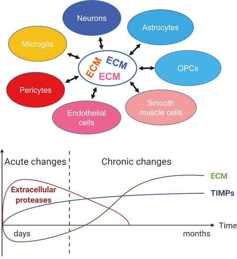
Multiple cells types release extracellular proteases and ECM molecules that assemble in diverse forms of ECM with multiple functional roles (upper panel). In brain diseases, a biphasic regulation of ECM is common: acute elevation of extracellular proteases leads to net ECM degradation, whereas a long-term increase in TIMP and ECM expression causes chronic ECM accumulation (lower panel). Abbreviations: ECM, extracellular matrix; OPCs, oligodendrocyte precursor cells; TIMP, tissue inhibitor of metalloproteinases. The lower panel was made using Biorender graphic software (https://biorender.com).
Integrin adhesion in brain assembly: From molecular structure to neuropsychiatric disorders
- Pages: 3831-3850
- First Published: 12 June 2020
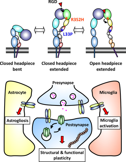
Integrins are extracellular matrix receptors mediating biochemical and mechanical signals at the cell surface. In the brain, they regulate development and function of neurons and glial cells. Defects in integrin expression or signaling contribute to autism spectrum disorder, epilepsy, schizophrenia, addiction, depression and Alzheimer's disease. We review how genetics and structure of integrins affect synaptic physiology and brain pathology, and discuss integrin-targeting strategies for therapies in neuropsychiatric disorders.
Mechanobiology of the brain in ageing and Alzheimer's disease
- Pages: 3851-3878
- First Published: 30 April 2020
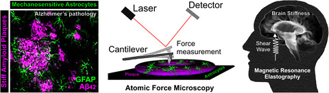
The mechanical properties of brain tissue can influence cell migration, neurogenesis, neurite outgrowth, regeneration and neuronal excitability. The extracellular microenvironment of neurons and glia changes in neurodegenerative disease states and, to a lesser extent, with “healthy ageing.” Understanding how perturbed mechanotransduction signalling influences the biochemistry and physiology of neurons and glia may reveal new drug targets for neurodegenerative disorders.
Extracellular matrix remodeling with stress and depression: Studies in human, rodent and zebrafish models
- Pages: 3879-3888
- First Published: 16 July 2020
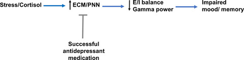
Stress and cortisol increase PNN levels. The PNN is a specialized form of ECM that is predominantly localized to PV-expressing GABAergic interneurons. Because increased PNN levels can enhance PV-expressing neuron-mediated inhibition of pyramidal cells, stress-associated increases in PNN deposition likely reduce E/I balance and gamma power to negatively affect mood and memory. Successful antidepressant therapy can counteract stress-related changes in PNN levels.
SPECIAL ISSUE ARTICLES
Aggrecan modulates the expression and phosphorylation of tau in a novel bigenic TauP301L - Acan mouse model
- Pages: 3889-3904
- First Published: 01 August 2020
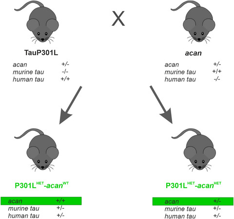
Perineuronal nets (PNs) are the most obvious manifestation of extracellular matrices in the brain. PNs carry neuroprotective functions as PN-ensheathed neurons are spared of tau pathology. To investigate the proposed association between PNs and Alzheimer’s disease, we generated a bigenic mouse model of TauP301L mice and heterozygous aggrecan mice. Our results highlight a correlation between aggrecan and P301L mutation triggered tau expression and phosphorylation in our bigenic mouse model.
Behavioural effects of extracellular matrix protein Fras1 depletion in the mouse
- Pages: 3905-3919
- First Published: 25 April 2020
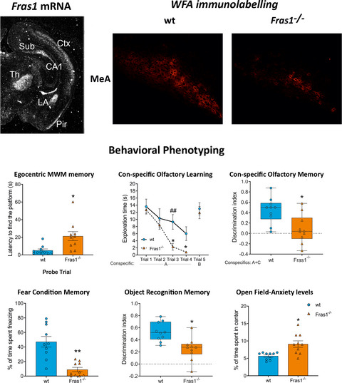
Fras1 is an extracellular protein, related to the Fraser syndrome. Fras1 mRNA is expressed in the choroid plexuses and in certain brain regions including cortical, hippocampal and amygdalar areas of mouse brain. Fras1−/− mice exhibit impaired egocentric spatial memory, aberrant olfactory learning and memory, reduced fear memory, recognition memory and anxiety, as well as disrupted extracellular matrix organization in cortical and subcortical areas as demonstrated by WFA labelling.
Poly I:C-induced maternal immune challenge reduces perineuronal net area and raises spontaneous network activity of hippocampal neurons in vitro
- Pages: 3920-3941
- First Published: 05 August 2020
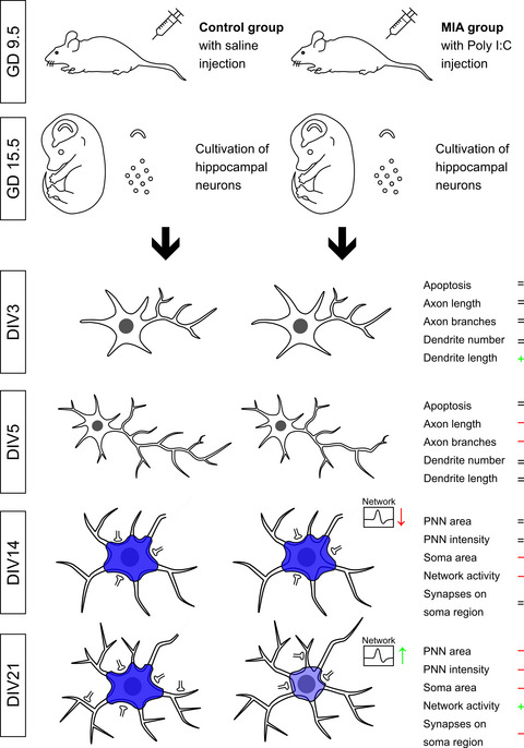
Pregnant BALB/c dams received Poly I:C injections at gestation day 9.5 in order to induce an activation of the maternal immune system. At gestation day 15.5 embryonic mice were isolated and hippocampi were prepared as well as digested to a single cell suspension. Morphological analysis of hippocampal neurons revealed alterations in axonal length and complexity. Most prominent abnormalities could be observed regarding perineuronal nets which were visualized via immunocytochemical Aggrecan staining. After 21 days in vitro hippocampal neurons displayed a reduced PNN area, staining intensity and a reduced soma area. These alterations were accompanied by a loss of excitatory synapse number on the neuronal soma. In addition, multielectrode array analysis unraveled a strong increase in spontaneous network activity at this point in time.
Neurocan genome-wide psychiatric risk variant affects explicit memory performance and hippocampal function in healthy humans
- Pages: 3942-3959
- First Published: 24 June 2020
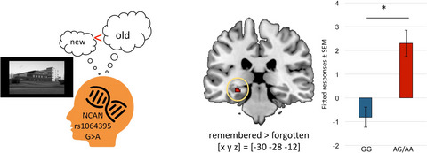
A genetic polymorphism in the vicinity of the NCAN gene has previously been associated with increased risk of schizophrenia and bipolar disorder. Here we investigated a potential association of this variant with memory function in healthy humans. Carriers of the risk allele exhibited lower verbal memory performance and atypically increased hippocampal activation during visual memory encoding. Furthermore, the risk allele was associated with reduced prefrontal grey matter density.
Molecular signature of extracellular matrix pathology in schizophrenia
- Pages: 3960-3987
- First Published: 18 October 2020
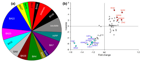
Growing evidence points to a critical involvement of the extracellular matrix (ECM) in the pathophysiology of schizophrenia (SZ). We tested the hypothesis that the expression of genes encoding for ECM molecules may be broadly disrupted in SZ across several cortical and subcortical brain regions. Gene expression profiling of 14 neocortical brain regions, caudate, putamen and hippocampus from control subjects and donors with SZ was conducted using Affymetrix microarray analysis. Analysis across brain regions revealed widespread dysregulation of ECM gene expression in cortical and subcortical brain regions in SZ. In (a) proportion of ECM-related differentially expressed genes (DEG) in SZ by the brain regions. The superior temporal cortex (BA22) showed the highest number of DEGs. In (b) ECM-related DEGs in SZ compared to UC across all of the analysed regions (blue-downregulated; red-upregulated). DEG's t-score (Y axis) values plotted against fold change (X axis).
Fine structure analysis of perineuronal nets in the ketamine model of schizophrenia
- Pages: 3988-4004
- First Published: 08 June 2020
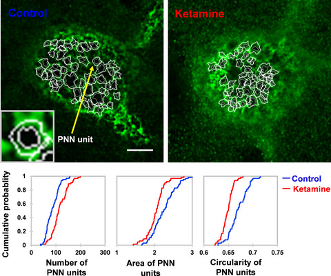
The neural extracellular matrix plays an important role in shaping synapses in healthy and diseased brains but the methods for quantitative analysis of its fine organization are just emerging. Here, we developed a tool for semi-automatic decomposition of perineuronal nets into a set of perisomatic circular-like units enriched in the perisynaptic extracellular matrix. We used this tool to reveal remarkable alterations in the number, size, and shape of these units in the ketamine model of schizophrenia.
Modified adeno-associated virus targets the bacterial enzyme chondroitinase ABC to select mouse neuronal populations in vivo using the Cre-LoxP system
- Pages: 4005-4015
- First Published: 21 November 2020
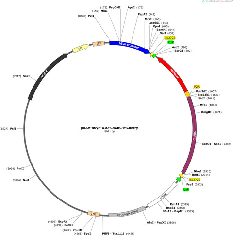
Cre-Dependent AAV-ChABC Construct: To express chondroitinaseABC (ChABC) in a Cre-recombinase dependent way, a shuttle plasmid containing a double floxed mCherry under the control of human synapsin promoter was used for the viral vector backbone, pAAV-hSyn-DIO-mCherry. We tested this virus in mice expressing a tamoxifen-regulated Cre recombinase in hippocampal area CA2.fx1
Bioinformatic analysis of the human brain extracellular matrix proteome in neurodegenerative disorders
- Pages: 4016-4033
- First Published: 20 May 2021
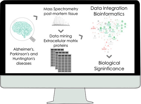
Schematic representation of the methodology used in the present study. The diagram illustrates the data mining of extracellular matrix proteins found altered in different proteomics studies that used post-mortem brain tissue from Alzheimer’s, Parkinson’s, and Huntington’s diseases patients followed by the creation of functional networks using the Cytoscape software.
Construction of a 3D brain extracellular matrix model to study the interaction between microglia and T cells in co-culture
- Pages: 4034-4050
- First Published: 20 September 2020
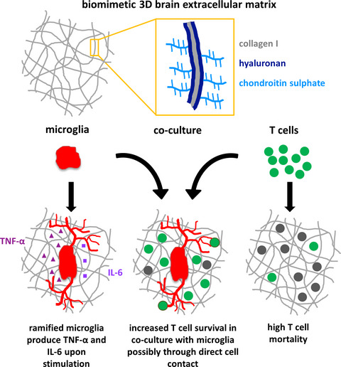
We developed a biomimetic 3D in vitro culture system for co-cultivation of microglia and T cells, where defined 3D collagen I-based matrices were modified with hyaluronan or chondroitin sulphate, resembling aspects of brain extracellular matrix. Murine microglia exhibited in vivo-like morphology and T cells showed enhanced survival when co-cultured with microglia. Microglia could be activated on the matrices by lipopolysaccharide resulting in interleukin-6 and tumour necrosis factor-α secretion.
Sign up for email alerts
Tools
Journal of FENS, published in association with Wiley
More from this journal
- Special Issues
- Trailblazers of Neuroscience
- Profiles of Women in Science
- Diversity Matters
- FENS-Kavli Network Editorials
- Nobel Prize Winners
- Call for papers
- Featured Articles
- EJN's Transparent Peer Review
- Aims & Scope in Chinese
- Chinese Webinar: How to Publish in EJN
- EJN Peer Review Training Webinar