

Journal list menu

Export Citations
Download PDFs
Cover Image
Cover Image
- Page: i
- First Published: 12 December 2023
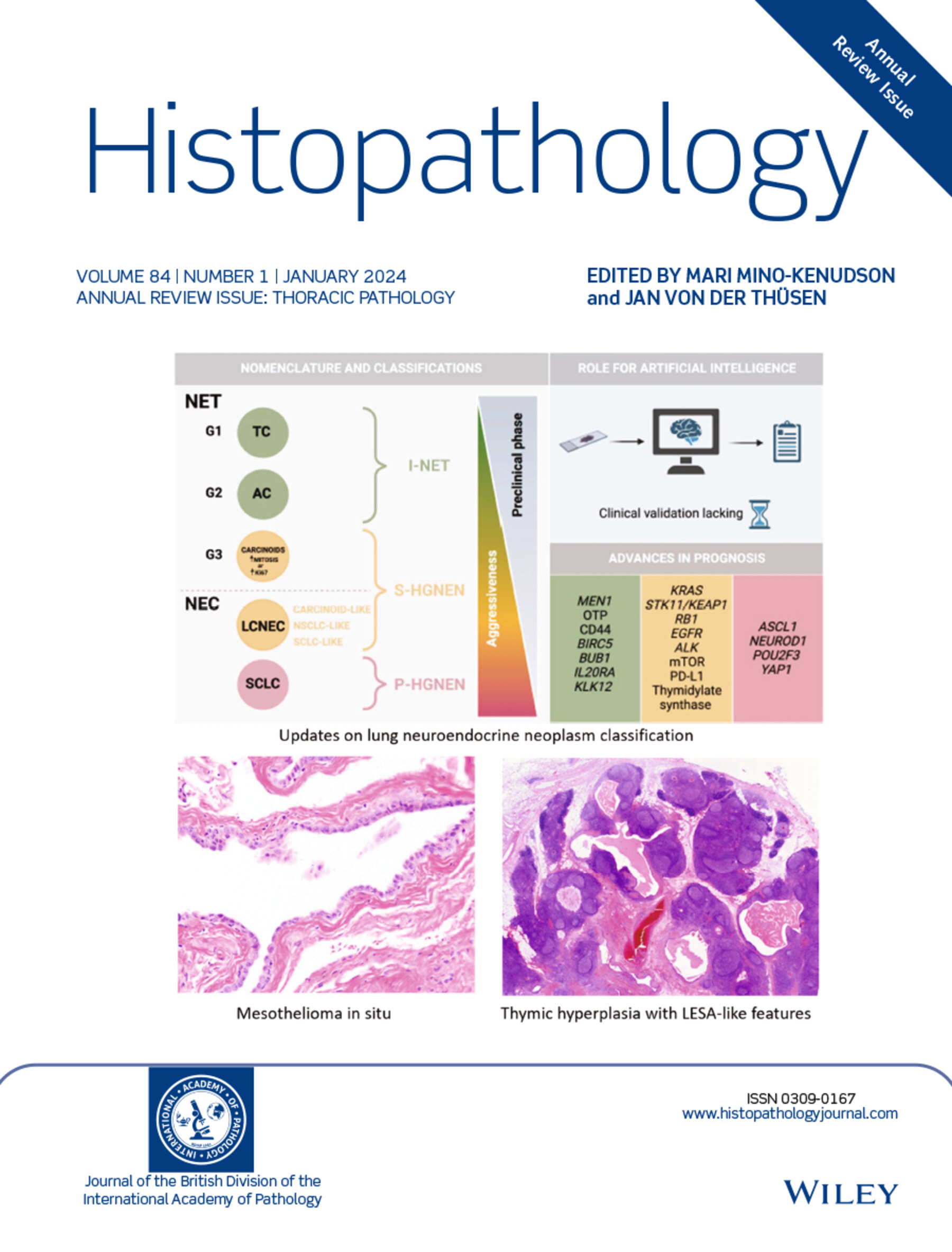
The cover image is based on the Review New developments in mesothelial pathology by Andrew Churg, https://doi.org/10.1111/his.15007; the Review Updates on lung neuroendocrine neoplasm classification by Giulia Vocino Trucco et al., https://doi.org/10.1111/his.15058; and the Review Benign lesions of the mediastinum by Tiemo Sven Gerber et al., https://doi.org/10.1111/his.15088.
Issue Information
Editorial
Annual Review Issue
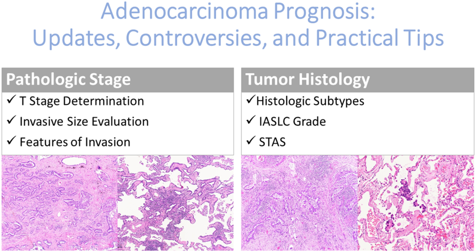
Updates on lung adenocarcinoma: invasive size, grading and STAS
- Pages: 6-17
- First Published: 23 October 2023
Pulmonary invasive mucinous adenocarcinoma
- Pages: 18-31
- First Published: 22 October 2023
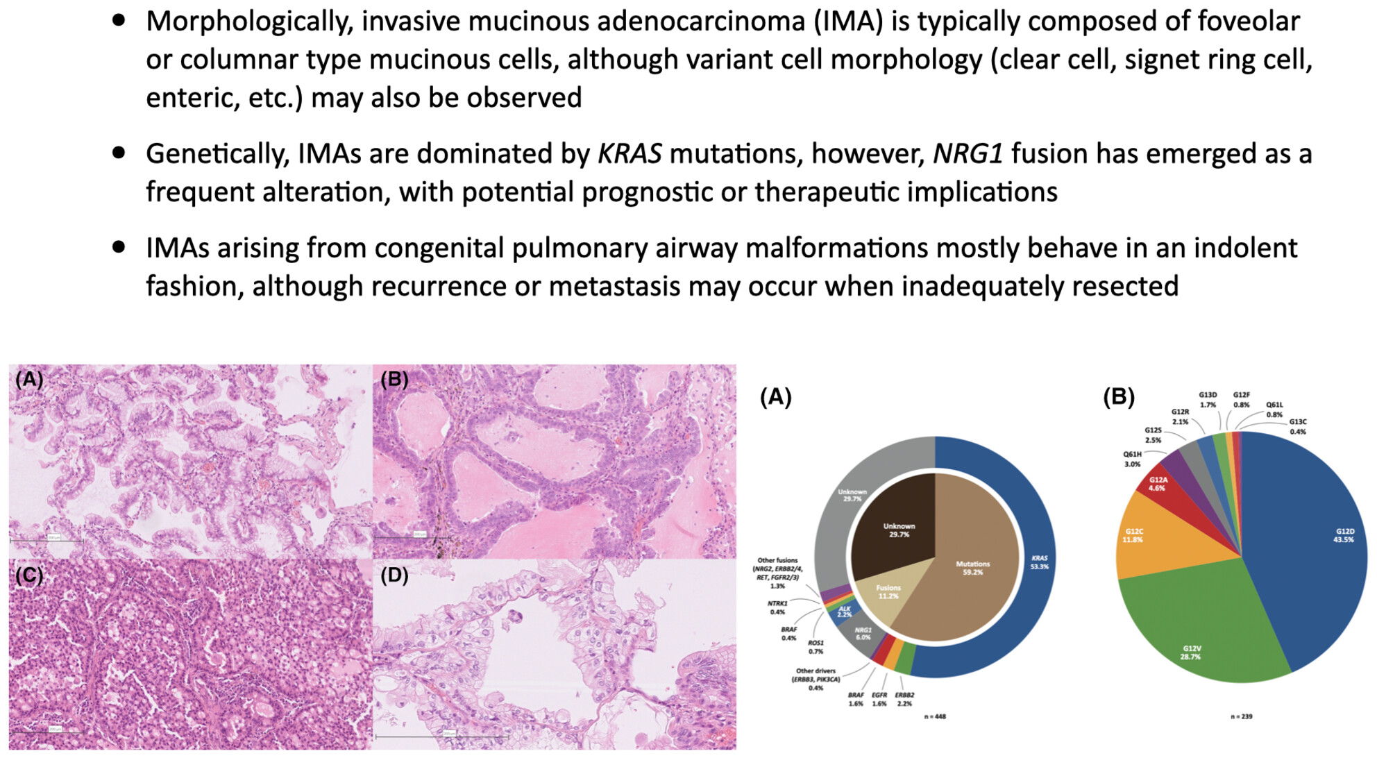
Classical and variant morphology of invasive mucinous adenocarcinoma.
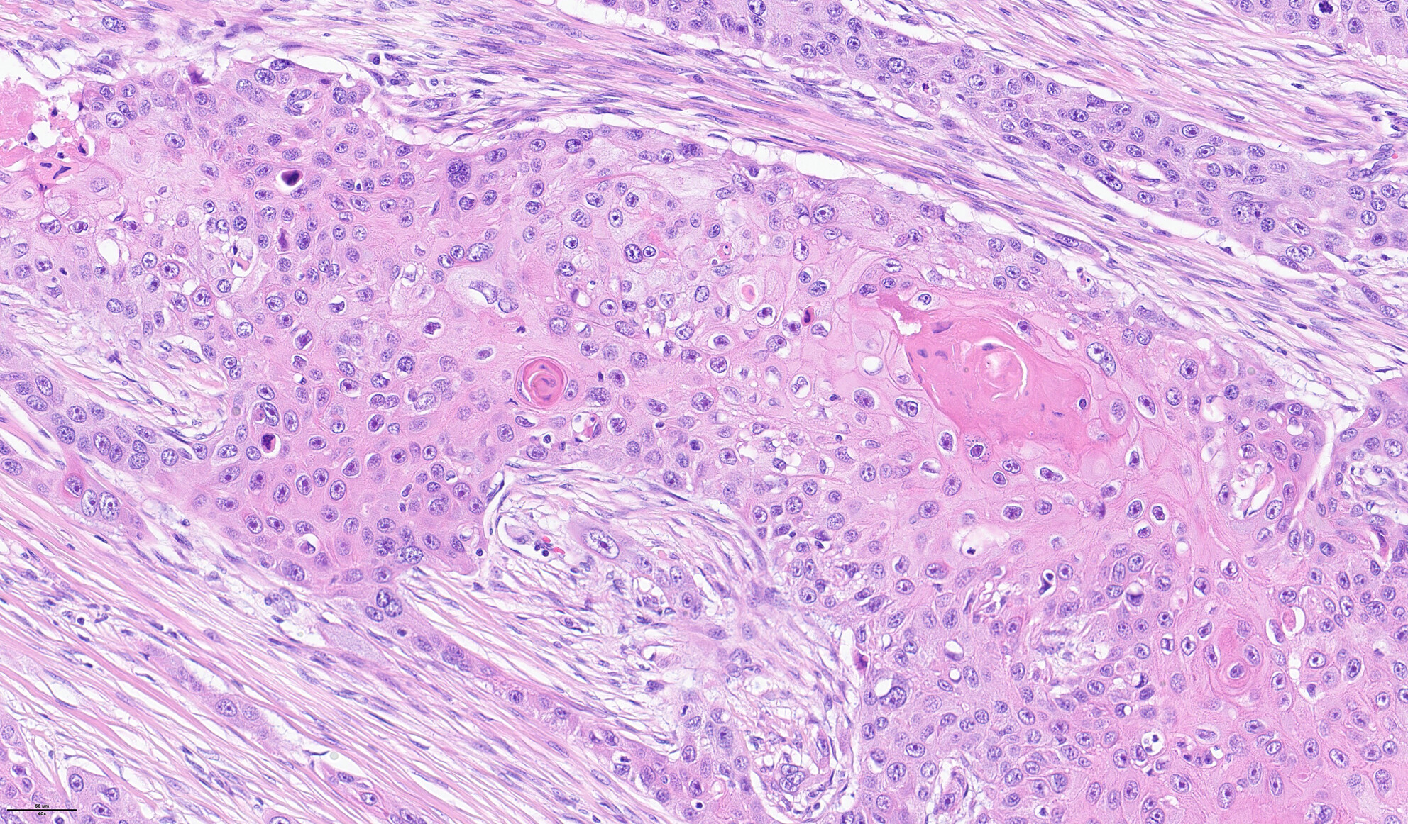
Pulmonary squamous cell carcinoma and lymphoepithelial carcinoma – morphology, molecular characteristics and differential diagnosis
- Pages: 32-49
- First Published: 07 November 2023
Molecular pathology of non-small cell carcinoma
- Pages: 50-66
- First Published: 07 November 2023
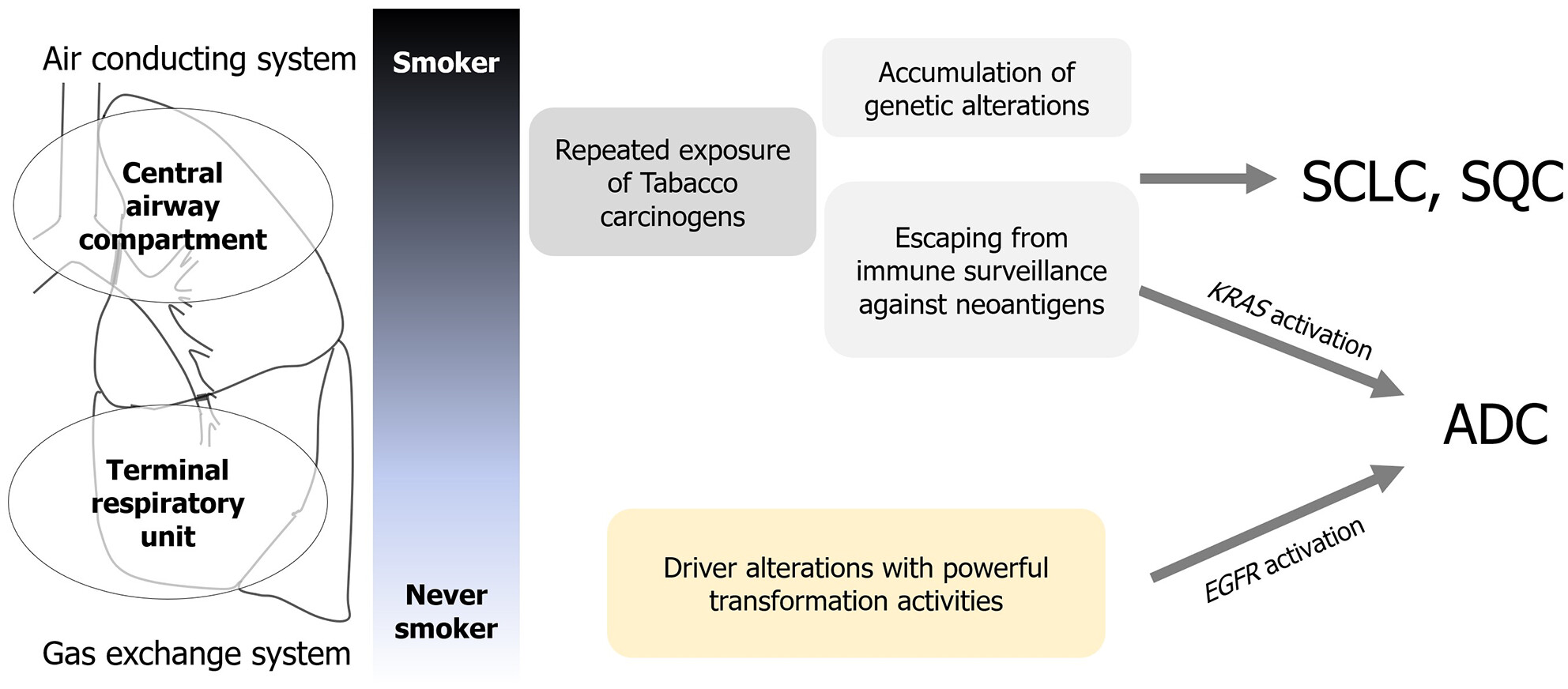
Two-compartment model in the putative molecular pathogenesis of lung cancer. The accumulation of genetic alterations with escaping immune surveillance is a key factor for tumours from the air-conducting system under the strong influence of tobacco smoke, whereas oncogene-addicted adenocarcinomas are driven by a single oncogenic mutation with powerful transformation activity.
Updates on lung neuroendocrine neoplasm classification
- Pages: 67-85
- First Published: 04 October 2023
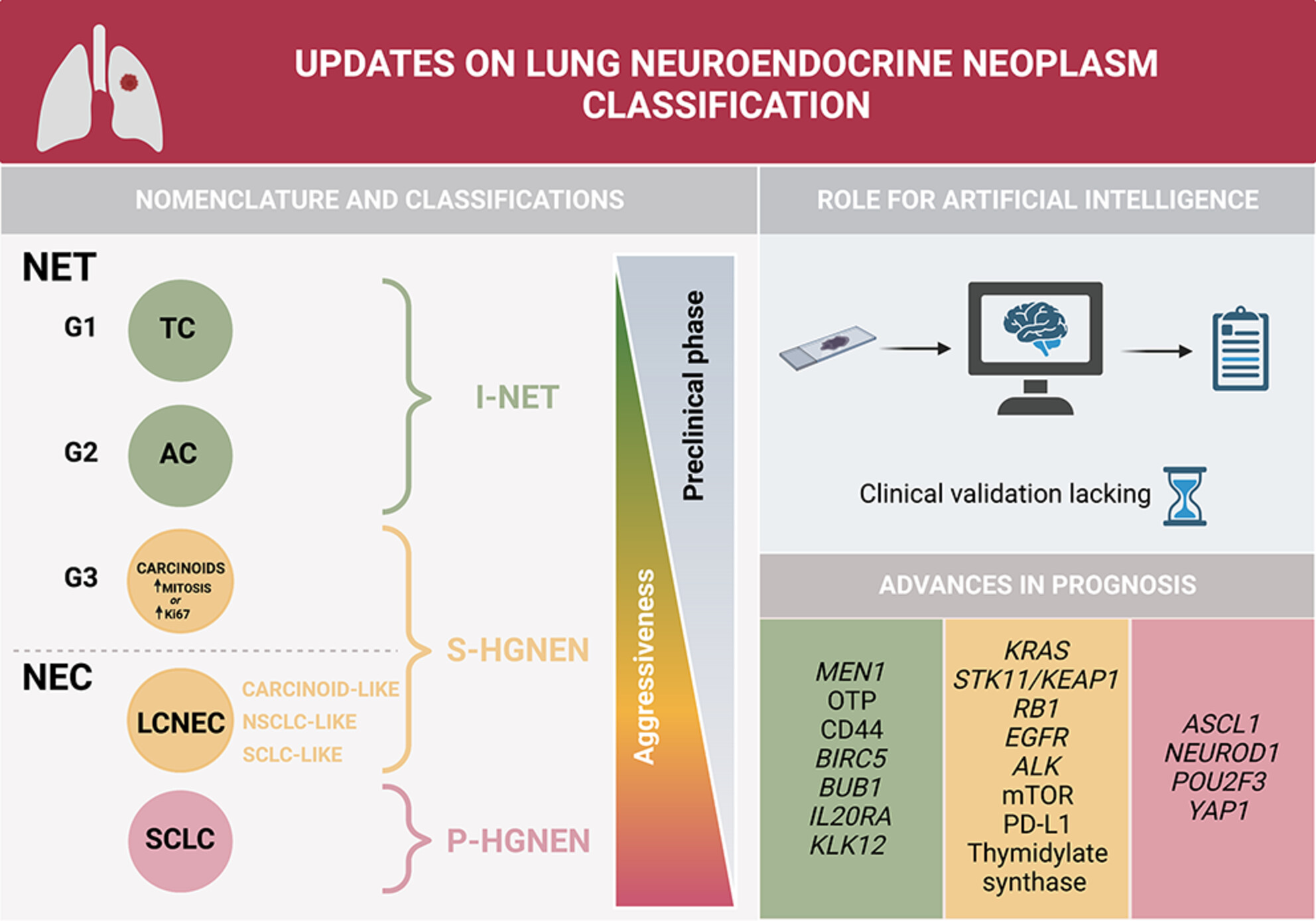
Highlights of major advances in lung neuroendocrine neoplasm classification, including the introduction of NET G3 in the classification framework, the potential impact of Artificial Intellignce, and the identification of novel prognostic molecular biomarkers.
NUT carcinoma and thoracic SMARCA4-deficient undifferentiated tumour: facts and controversies
- Pages: 86-101
- First Published: 24 October 2023
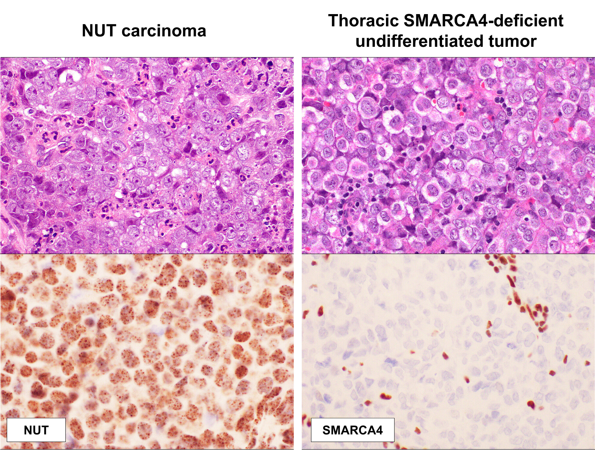
This review provides an update on NUT carcinoma and thoracic SMARCA4-deficient undifferentiated tumor, including their key clinicopathological/molecular characteristics and controversies.
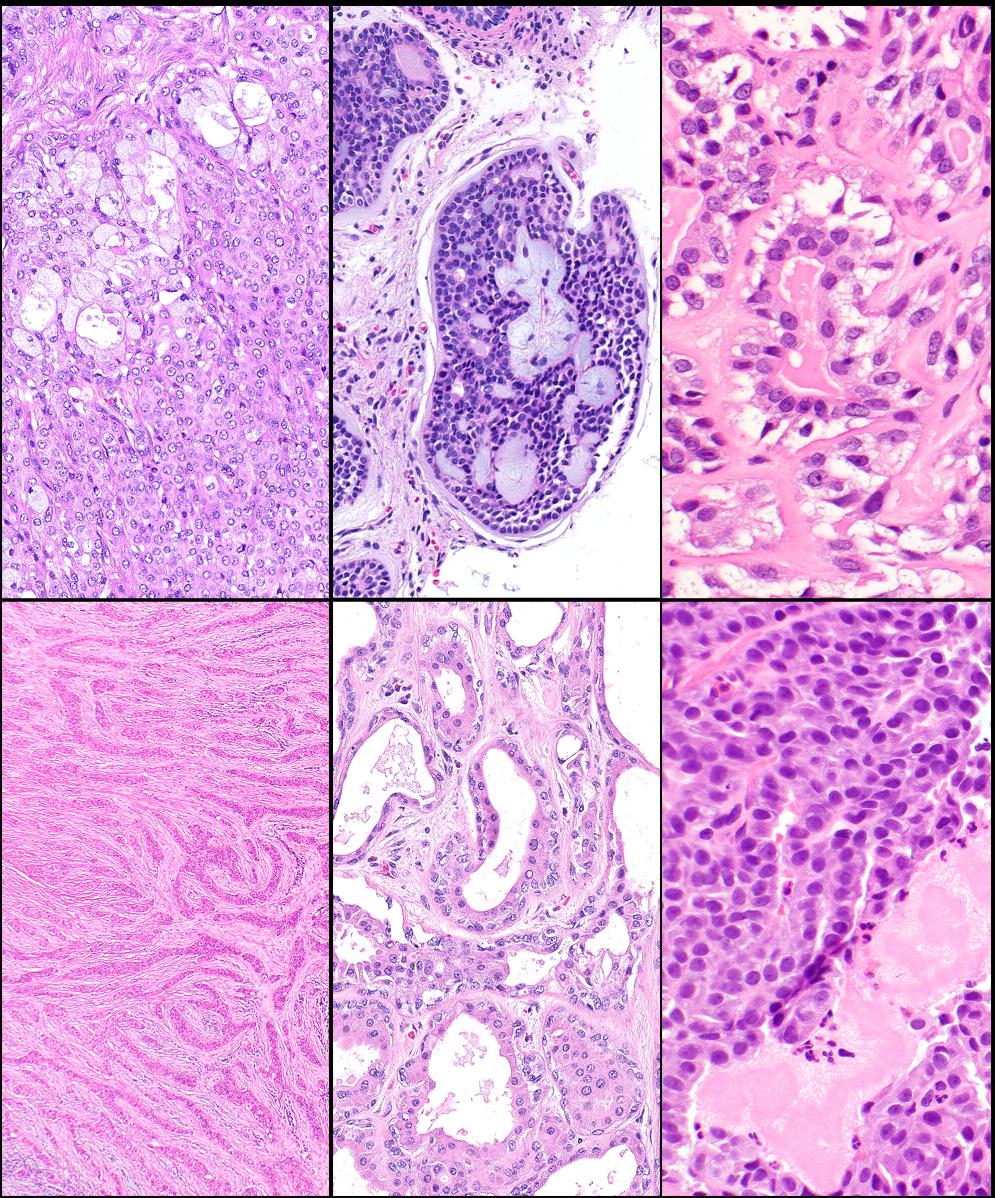
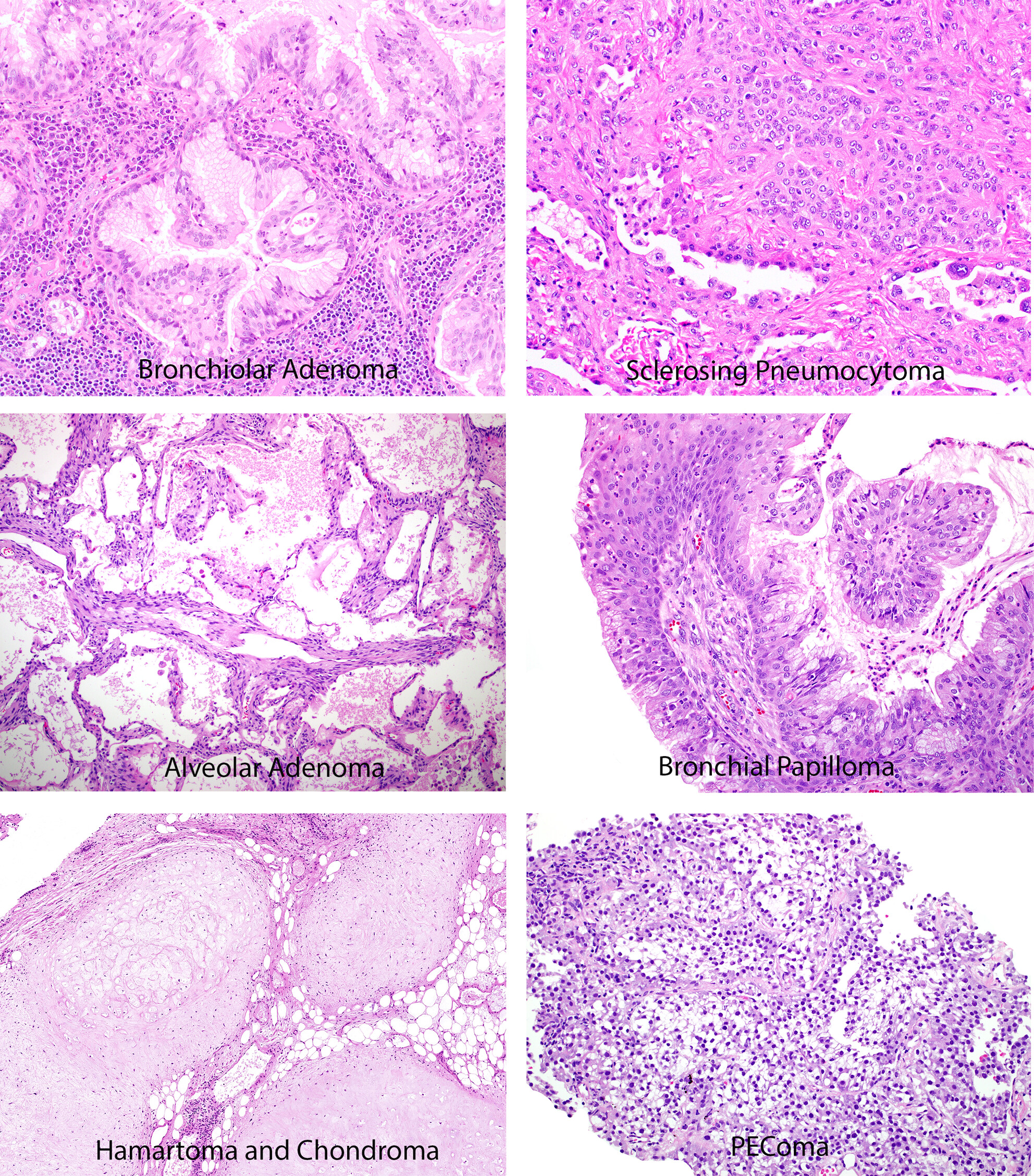
Recent developments in the pathology of primary pulmonary salivary gland-type tumours
- Pages: 102-123
- First Published: 11 September 2023
New developments in mesothelial pathology
- Pages: 136-152
- First Published: 11 September 2023
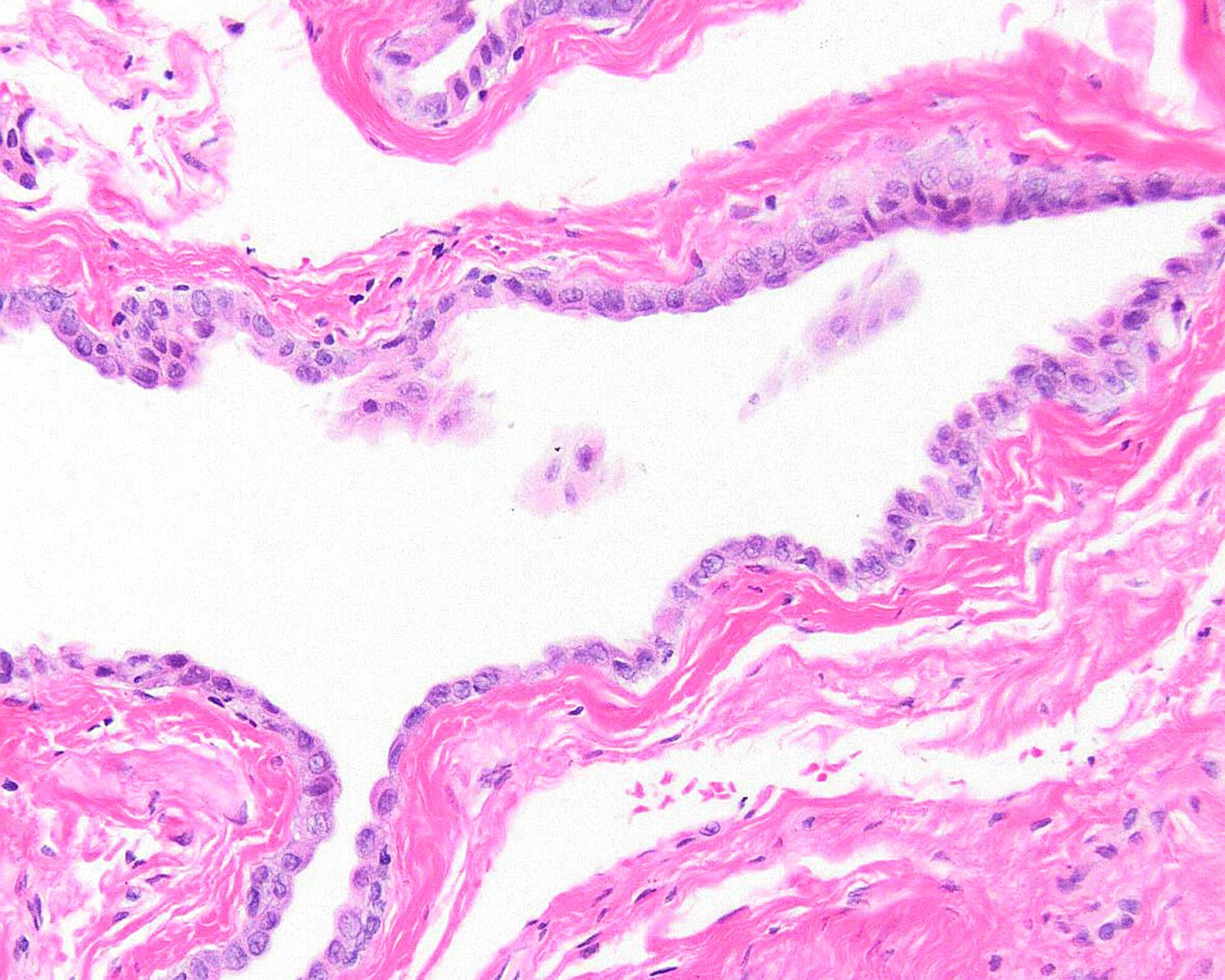
An example of mesothelioma in situ, a new entity discussed in this review.
Updates on grading mesothelioma
- Pages: 153-162
- First Published: 23 October 2023
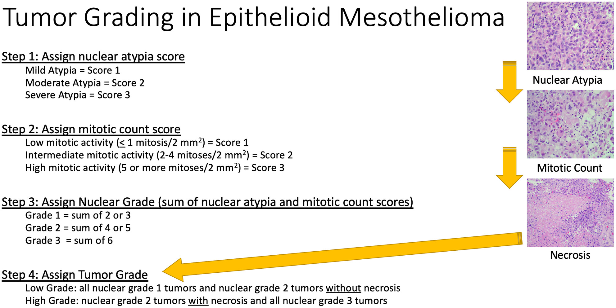
Grading of epithelioid mesothelioma is now recommended and is a powerful prognostic tool. Grading requires calculation of an atypia score and mitotic count score which yields a nuclear grade that is combined with the presence or absence of necrosis to determine overall grade (low or high).
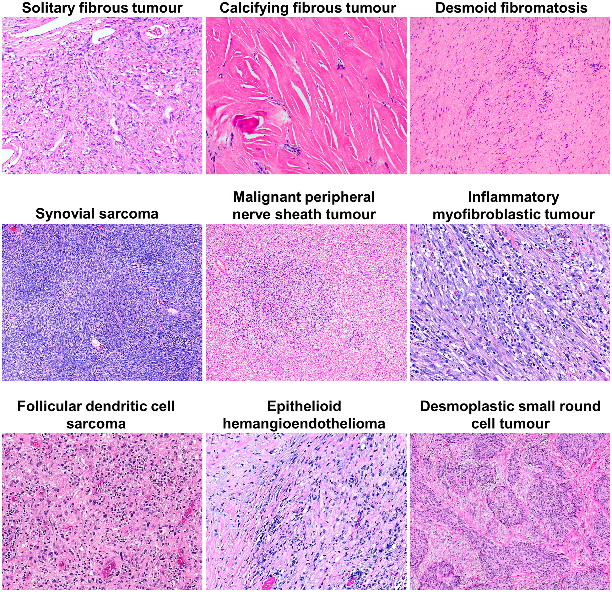
Mesenchymal tumours of the pleura: review and update
- Pages: 163-182
- First Published: 10 September 2023
Benign lesions of the mediastinum
- Pages: 183-195
- First Published: 21 November 2023
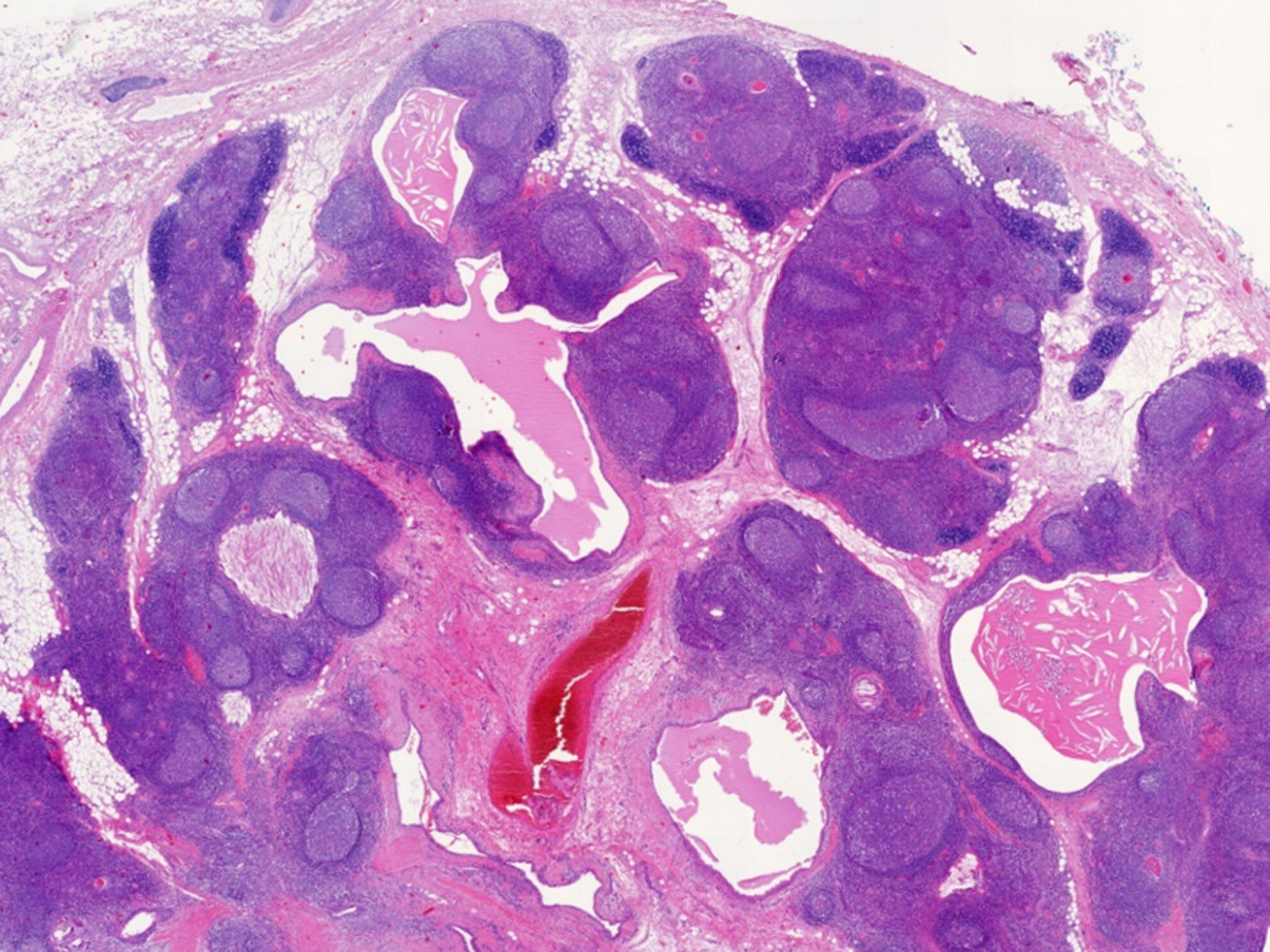
Thymic hyperplasia with lymphoepithelial sialadenitis (LESA)-like features as an example of a thymic tumorous lesion showing an association with non-myasthenic autoimmune diseases and lymphomas.
Thymic epithelial tumours: histopathological classification and differential diagnosis
- Pages: 196-215
- First Published: 23 November 2023
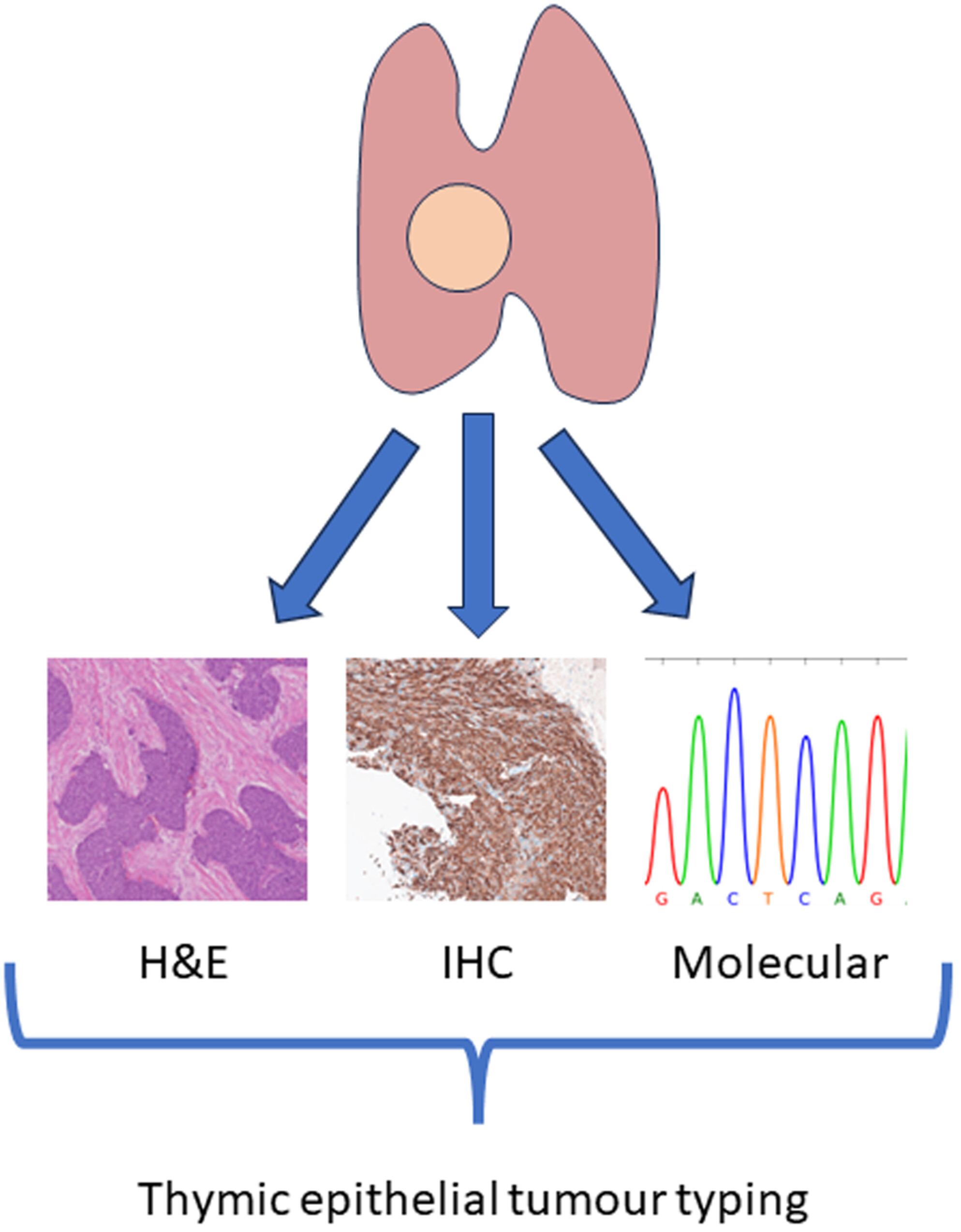
In this review, the various subtypes of thymic epithelial tumours are described, with attention to differential diagnosis and ancillary techniques, as well as limitations of biopsy and frozen section diagnosis, and the clinical relevance for prognosis and treatment.
Primary germ cell tumours of the mediastinum: A review with emphasis on diagnostic challenges
- Pages: 216-237
- First Published: 23 November 2023
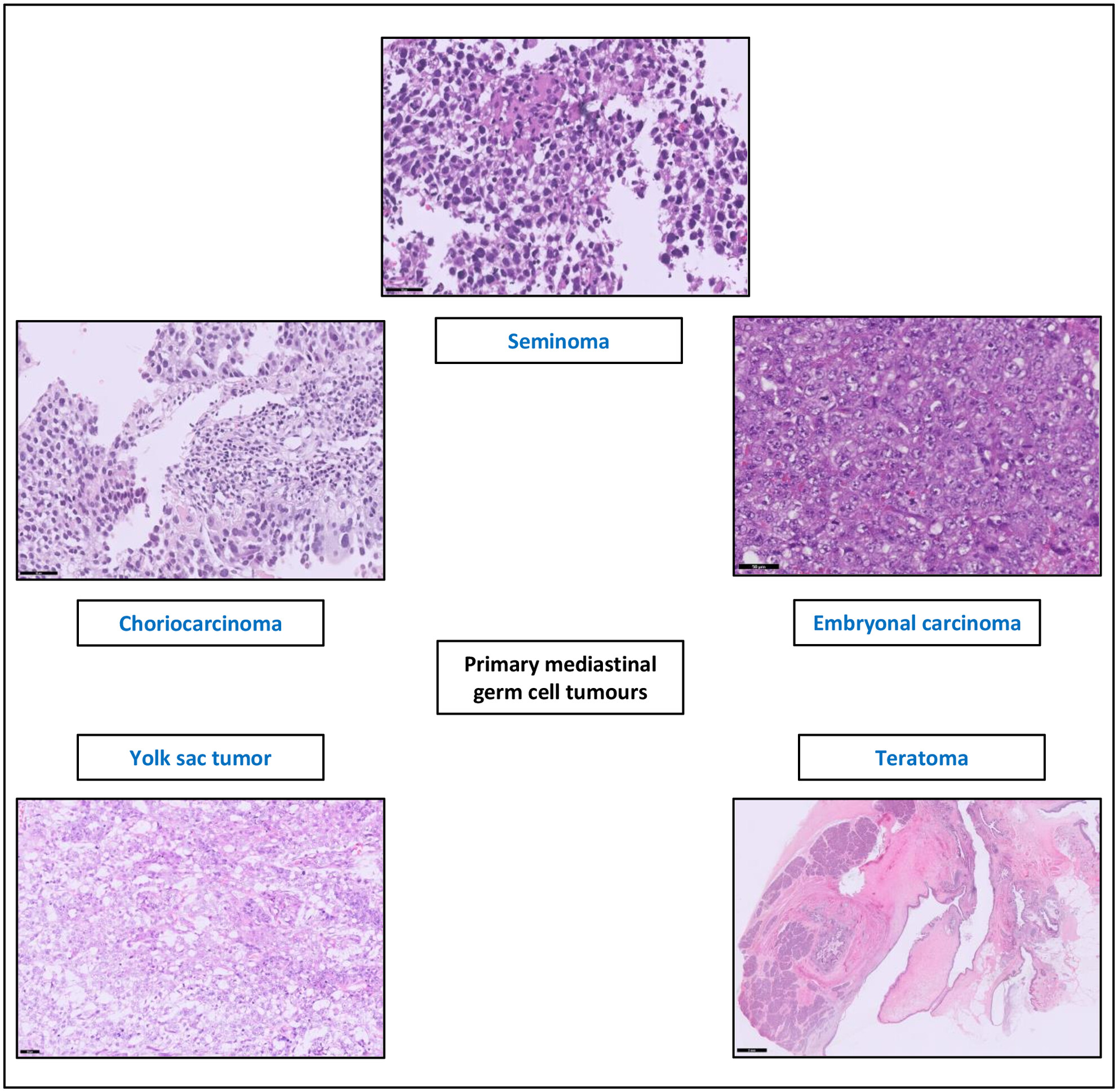
Primary mediastinal germ cell tumours encompass five histological subtypes: seminoma, embryonal carcinoma, teratoma, yolk-sac tumour and choriocarcinoma. Seminomas consist of medium-sized cells with clear to eosinophilic cytoplasm and prominent nucleoli. Embryonal carcinomas with solid growth pattern, sometimes glandular or papillary growth pattern, contain pleomorphic cells with overlapping nuclei and prominent nucleoli. Teratomas can show differentiation of all three germ cell layers, usually forming cysts lined by squamous, respiratory, or intestinal epithelium. Solid areas contain pancreatic tissue, fat tissue, and smooth muscles. Yolk-sac tumours can show different growth patterns with reticular growth pattern as the most common. Choriocarcinomas consist of bilamellar structures of mononucleated cytotrophoblasts and multinucleated syncytiotrophoblasts.
Neurogenic tumours of the posterior mediastinum and differential diagnosis considerations
- Pages: 238-252
- First Published: 28 September 2023
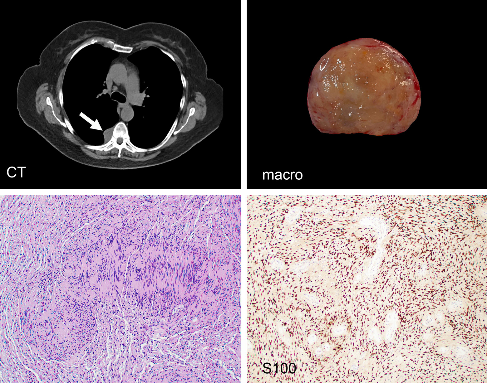
Schwannoma of the posterior mediastinum. This is the most common neural tumour of the mediastinum. The gross and microscopic aspect is similar in every respect to Schwannoma's of soft tissue elsewhere.
Sign up for email alerts
Tools
Published on behalf of the British Division of the International Academy of Pathology