

Journal list menu

 Issue
IssueJournal of Comparative Neurology: Volume 530, Issue 18
3105-3287December 2022
Export Citations
Download PDFs
ISSUE INFORMATION - TOC
The Journal of Comparative Neurology, Table of Content, Vol. 530, No. 18, December, 2022
- Page: 3105
- First Published: 19 October 2022
RESEARCH ARTICLES
Comparative analysis of astrocytes in the prefrontal cortex of primates: Insights into the evolution of human brain energetics
- Pages: 3106-3125
- First Published: 20 July 2022
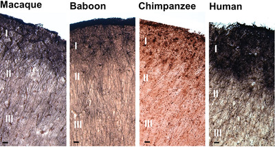
We quantified astrocytes expressing glial fibrillary acidic protein in human and nonhuman primates. We report that astrocyte density, soma volume, and ratio of astrocytes to total glia cells in layer I of Brodmann.s area 9 increased significantly with brain size enlargement and was greatest in humans. Roman numerals denote cortical layer. Scale bars = 25 µm.
3D-atlas of the brain of the cockroach Rhyparobia maderae
- Pages: 3126-3156
- First Published: 29 August 2022

Cockroaches are model organisms for research on olfaction, spatial orientation and circadian control of behavior. To facilitate further studies on these topics, we provide a detailed 3D atlas of brain neuropils, fiber tracts, and commissures of the cockroach Rhyparobia maderae, which reveals novel insights into the organization of the cockroach brain.
Neuropeptide S (NPS) neurons: Parabrachial identity and novel distributions
- Pages: 3157-3178
- First Published: 29 August 2022
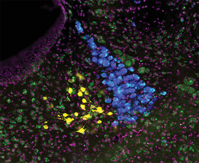
Neuropeptide S (NPS) neurons in the parabrachial region are a subset of Atoh1-derived, Foxp2-expressing neurons that do not express Pdyn, Calca, or Cck. We also identified novel populations of NPS neurons from Cre-reporter expression in the nucleus incertus, anterior hypothalamus, and lateral habenula.
The parabigeminal nucleus is a source of “retinogeniculate replacement terminals” in mice that lack retinofugal input
- Pages: 3179-3192
- First Published: 06 September 2022
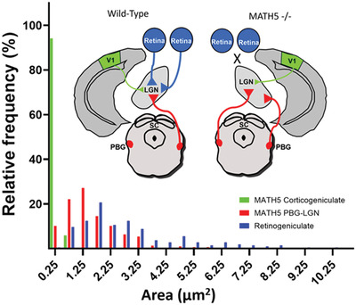
In the dorsal lateral geniculate nucleus (LGN) of mice that lack retinal input, a population of large terminals supplants the synaptic arrangements normally made by the missing retinogeniculate terminals. To identify potential sources of these “retinogeniculate replacement terminals,” we used mutant mice (math5–/–) which lack retinofugal projections due to the failure of retinal ganglion cells to develop. In this line, we labeled LGN terminals that originate from the primary visual cortex (V1) or the parabigeminal nucleus (PBG), and compared their ultrastructure to retinogeniculate, V1, or PBG terminals in the LGN of C57Blk6 (WT) mice (schematically depicted above graph). Corticogeniculate terminals labeled in WT and math5–/– mice were similar in size and both groups were significantly smaller than WT retinogeniculate terminals. In contrast, the PBG projection in math5–/– mice was extensive and there was considerable overlap in the sizes of retinogeniculate terminals in WT mice and PBG terminals in math5–/– mice (summarized in histogram). The data indicate that V1 is not a source of “retinogeniculate replacement terminals” and suggest that large PBG terminals expand their innervation territory to replace retinogeniculate terminals in their absence.
Transient neurochemical features of the perigeniculate neurons during early postnatal development of the cat
- Pages: 3193-3208
- First Published: 29 August 2022
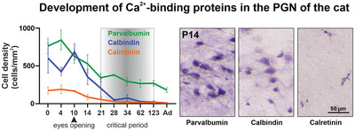
Development of Ca2+-binding proteins in the PGN of the cat.
Quantification of CGRP-immunoreactive myenteric neurons in mouse colon
- Pages: 3209-3225
- First Published: 31 August 2022
Calcitonin gene-related peptide (CGRP) is a putative marker of multiaxonal, intrinsic primary afferent neurons in mouse colon. This study quantifies the proportion of myenteric nerve cell bodies along the mouse colon that are immunoreactive to CGRP.
Diversity in spatial frequency, temporal frequency, and speed tuning across mouse visual cortical areas and layers
- Pages: 3226-3247
- First Published: 07 September 2022
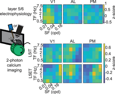
Using extracellular electrophysiology and two-photon calcium imaging genetically targeted to different layer 5 cell classes, we characterize the spatial frequency (SF), temporal frequency (TF), and speed tuning of deep (layer 5/6) cortical neurons in mouse primary visual cortex (V1) and higher visual cortical areas anterolateral (AL) and posteromedial (PM). We find that deep-layer neurons, like superficial layer neurons, are functionally specialized for different spatial and temporal frequencies in different visual areas. However, we also found a larger range of preferred SFs and TFs than previously reported in superficial cortical layers. We also find that direction tuning is much more prominent for layer 5 extratelencephalically than layer 5 intratelencephalically projecting neurons in multiple visual areas.
Tonotopic differentiation of presynaptic neurotransmitter-releasing machinery in the auditory brainstem during the prehearing period and its selective deficits in Fmr1 knockout mice
- Pages: 3248-3269
- First Published: 06 September 2022
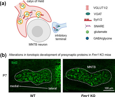
We identified protein-specific and age-dependent tonotopic distributions of five key presynaptic proteins in the developing auditory brainstem and their selective alterations in Fmr1 KO mice. The tonotopic maturation of the presynaptic machinery may provide a potential mechanism for tonotopic differentiation of neurotransmission during normal and abnormal development of auditory circuits.
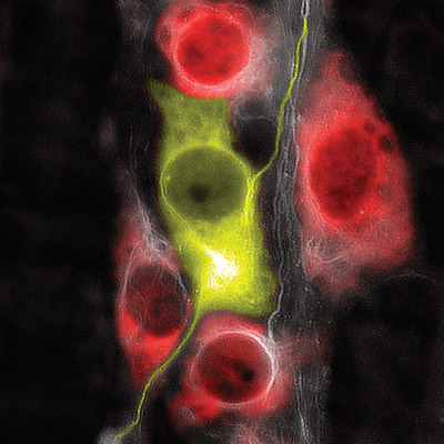
Quantitative spatial analysis reveals that the local axons of lamina I projection neurons and interneurons exhibit distributions that predict distinct roles in spinal sensory processing
- Pages: 3270-3287
- First Published: 12 September 2022
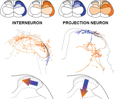
Quantitative analyses of spinal lamina I projection neurons (PNs) and interneurons (INs) revealed almost identical dendritic patterns but local axon collateral distribution patterns that are distinct. INs mostly establish connections within the superficial dorsal horn and may bridge its medial and lateral halves. PNs also relay their input locally, to deeper laminae of the spinal gray matter and may link other ascending systems and participate directly in the nociceptive withdrawal reflex.