

Integrative microendoscopic system combined with conventional microscope for live animal tissue imaging
Martin Köhler
The Rolf Luft Research Center for Diabetes and Endocrinology, Karolinska Institutet, Stockholm, Sweden
Search for more papers by this authorBjorn Paulson
Institute of Physics and Applied Physics, Yonsei University, Seoul, Korea
Biomedical Engineering Research Center, Asan Institute for Life Science, Asan Medical Center, Seoul, Korea
Search for more papers by this authorYoungkyu Kim
Biomedical Engineering Research Center, Asan Institute for Life Science, Asan Medical Center, Seoul, Korea
Search for more papers by this authorSanghwa Lee
Biomedical Engineering Research Center, Asan Institute for Life Science, Asan Medical Center, Seoul, Korea
Search for more papers by this authorAndrea Dicker
The Rolf Luft Research Center for Diabetes and Endocrinology, Karolinska Institutet, Stockholm, Sweden
Search for more papers by this authorPim van Krieken
The Rolf Luft Research Center for Diabetes and Endocrinology, Karolinska Institutet, Stockholm, Sweden
Search for more papers by this authorJae Young Kim
Research Institute for Skin Imaging, Korea University Medical Center, Seoul, Korea
Search for more papers by this authorChan-Gi Pack
Department of Convergence Medicine, University of Ulsan, College of Medicine, Seoul, Korea
Biomedical Science Research Center, Asan Institute for Life Sciences, Asan Medical Center, Seoul, Korea
Search for more papers by this authorJinmyoung Joo
Biomedical Engineering Research Center, Asan Institute for Life Science, Asan Medical Center, Seoul, Korea
Department of Convergence Medicine, University of Ulsan, College of Medicine, Seoul, Korea
Search for more papers by this authorCorresponding Author
Per-Olof Berggren
The Rolf Luft Research Center for Diabetes and Endocrinology, Karolinska Institutet, Stockholm, Sweden
Correspondence
Per-Olof Berggren, The Rolf Luft Research Center for Diabetes and Endocrinology, Karolinska Institutet, Stockholm SE-17176, Sweden.
Email: [email protected]
Jun Ki Kim, Biomedical Engineering Research Center, Asan Institute for Life Science, Asan Medical Center, 88, Olympic-ro 43-gil, Songpa-gu, Seoul 05505, Korea.
Email: [email protected]
Search for more papers by this authorCorresponding Author
Jun Ki Kim
Biomedical Engineering Research Center, Asan Institute for Life Science, Asan Medical Center, Seoul, Korea
Department of Convergence Medicine, University of Ulsan, College of Medicine, Seoul, Korea
Correspondence
Per-Olof Berggren, The Rolf Luft Research Center for Diabetes and Endocrinology, Karolinska Institutet, Stockholm SE-17176, Sweden.
Email: [email protected]
Jun Ki Kim, Biomedical Engineering Research Center, Asan Institute for Life Science, Asan Medical Center, 88, Olympic-ro 43-gil, Songpa-gu, Seoul 05505, Korea.
Email: [email protected]
Search for more papers by this authorMartin Köhler
The Rolf Luft Research Center for Diabetes and Endocrinology, Karolinska Institutet, Stockholm, Sweden
Search for more papers by this authorBjorn Paulson
Institute of Physics and Applied Physics, Yonsei University, Seoul, Korea
Biomedical Engineering Research Center, Asan Institute for Life Science, Asan Medical Center, Seoul, Korea
Search for more papers by this authorYoungkyu Kim
Biomedical Engineering Research Center, Asan Institute for Life Science, Asan Medical Center, Seoul, Korea
Search for more papers by this authorSanghwa Lee
Biomedical Engineering Research Center, Asan Institute for Life Science, Asan Medical Center, Seoul, Korea
Search for more papers by this authorAndrea Dicker
The Rolf Luft Research Center for Diabetes and Endocrinology, Karolinska Institutet, Stockholm, Sweden
Search for more papers by this authorPim van Krieken
The Rolf Luft Research Center for Diabetes and Endocrinology, Karolinska Institutet, Stockholm, Sweden
Search for more papers by this authorJae Young Kim
Research Institute for Skin Imaging, Korea University Medical Center, Seoul, Korea
Search for more papers by this authorChan-Gi Pack
Department of Convergence Medicine, University of Ulsan, College of Medicine, Seoul, Korea
Biomedical Science Research Center, Asan Institute for Life Sciences, Asan Medical Center, Seoul, Korea
Search for more papers by this authorJinmyoung Joo
Biomedical Engineering Research Center, Asan Institute for Life Science, Asan Medical Center, Seoul, Korea
Department of Convergence Medicine, University of Ulsan, College of Medicine, Seoul, Korea
Search for more papers by this authorCorresponding Author
Per-Olof Berggren
The Rolf Luft Research Center for Diabetes and Endocrinology, Karolinska Institutet, Stockholm, Sweden
Correspondence
Per-Olof Berggren, The Rolf Luft Research Center for Diabetes and Endocrinology, Karolinska Institutet, Stockholm SE-17176, Sweden.
Email: [email protected]
Jun Ki Kim, Biomedical Engineering Research Center, Asan Institute for Life Science, Asan Medical Center, 88, Olympic-ro 43-gil, Songpa-gu, Seoul 05505, Korea.
Email: [email protected]
Search for more papers by this authorCorresponding Author
Jun Ki Kim
Biomedical Engineering Research Center, Asan Institute for Life Science, Asan Medical Center, Seoul, Korea
Department of Convergence Medicine, University of Ulsan, College of Medicine, Seoul, Korea
Correspondence
Per-Olof Berggren, The Rolf Luft Research Center for Diabetes and Endocrinology, Karolinska Institutet, Stockholm SE-17176, Sweden.
Email: [email protected]
Jun Ki Kim, Biomedical Engineering Research Center, Asan Institute for Life Science, Asan Medical Center, 88, Olympic-ro 43-gil, Songpa-gu, Seoul 05505, Korea.
Email: [email protected]
Search for more papers by this authorAbstract
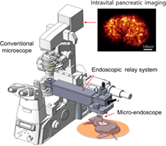
Intravital optical imaging technology is essential for minimally invasive optical diagnosis and treatment in small animal disease models. High-resolution imaging requires high-resolution optical probes, and high-resolution optical imaging systems based on highly precise and advanced technologies and therefore, associated with high-system costs. Besides, in order to acquire small animal live images, special types of animal imaging setups are indispensable. In this paper, a microendoscopic system is designed as an add-on to existing conventional imaging microscopes, reducing the price of complete confocal endomicroscopic systems. The proposed attachable system can be configured for confocal microscopes from common manufacturers and this enables users to acquire live animal cellular images from a conventional system. It features a 4f optical plane relay system, a rotary stage for side-view endoscopic probes, and an endoscopic probe mount which swings between the horizontal and the vertical. The system could be widely useful for biological studies of animal physiology and disease models.
Supporting Information
| Filename | Description |
|---|---|
| jbio201800206-sup-0101-author-biographies.docxapplication/docx, 81.9 KB | Author Biographies |
| jbio201800206-sup-0001-FigureS1.jpgJPEG image, 1 MB | Figure S1 Detailed cutaway of the device design, with a focus on the optical components. Components shown in blue are mirror mounts, and in black are lens mounts, which each immobilize two achromatic doublets in the optical path. The objective lens in green is held in the beam path and connected to the Z-stage by the microscope mount, shown in gold. |
| jbio201800206-sup-0002-FigureS2.jpgJPEG image, 778.3 KB | Figure S2 Engineering schematic of the probe mount, showing a semitransparent probe mounted in the v-groove, and immobilized by the screw clamp. |
| jbio201800206-sup-0003-FigureS3.jpgJPEG image, 938.9 KB | Figure S3 Engineering schematic of the objective mount, showing its connection to the z-stage and independence from the rotational stage. The entire objective mount and probe assembly swings with the swing mirror. |
| jbio201800206-sup-0004-FigureS4.jpgJPEG image, 1.2 MB | Figure S4 Modulation transfer function (MTF) analysis of the attachable relay system. (A) Image of a USAF 1951 target. (B) MTF as calculated from the slanted line method. |
| jbio201800206-sup-0005-MovieS1.avivideo/avi, 4.5 MB | Movie S1 Z-stack movie of the FITC injected murine vasculature (30um depth with 3um steps) |
Please note: The publisher is not responsible for the content or functionality of any supporting information supplied by the authors. Any queries (other than missing content) should be directed to the corresponding author for the article.
REFERENCES
- 1M. E. Bocarsly, W. Jiang, C. Wang, J. T. Dudman, N. Ji, Y. Aponte, Biomed. Opt. Express 2015, 6, 11.
- 2J. C. Jung, J. Neurophysiol. 2004, 92, 5.
- 3R. P. J. Barretto, S. Gillis-Smith, J. Chandrashekar, D. A. Yarmolinsky, M. J. Schnitzer, N. J. P. Ryba, C. S. Zuker, Nature 2014, 517, 7534.
- 4P. Kim, M. Puoris'haag, D. Coté, C. P. Lin, S. H. Yun, J. Biomed. Opt. 2008, 13, 1.
- 5P. Kim, E. Chung, H. Yamashita, K. E. Hung, A. Mizoguchi, R. Kucherlapati, D. Fukumura, R. K. Jain, S. H. Yun, Nat. Methods 2010, 7, 4.
10.1038/nmeth.f.315 Google Scholar
- 6J. K. Kim, W. M. Lee, P. Kim, M. Choi, K. Jung, S. Kim, S. H. Yun, Nat. Protoc. 2012, 7, 8.
- 7J. Knittel, L. Schnieder, G. Buess, B. Messerschmidt, T. Possner, Opt. Commun. 2001, 188, 5.
- 8A. R. Rouse, A. Kano, J. A. Udovich, S. M. Kroto, A. F. Gmitro, Appl. Optics 2004, 43, 31.
- 9K.-B. Sung, R. Richards-Kortum, M. Follen, A. Malpica, C. Liang, M. Descour, Opt. Express 2003, 11, 24.
- 10A. A. Tanbakuchi, J. A. Udovich, A. R. Rouse, K. D. Hatch, A. F. Gmitro, Am. J. Obstet. Gynecol. 2010, 202, 1.
- 11A. F. Gmitro, D. Aziz, Opt. Lett. 1993, 18, 8.
- 12W. Göbel, J. N. D. Kerr, A. Nimmerjahn, F. Helmchen, Opt. Lett. 2004, 29, 21.
- 13M. Kim, J. Hong, J. Kim, H. Shin, Biomed. Opt. Express 2017, 8, 6.
- 14B. Messerschmidt, G. Matz, Opt. Photonik 2014, 9, 4.
- 15J. K. Kim, J. Choi, S. H. Yun, OSA, 2011, p. FTuX3.
- 16J. K. Kim, J. W. Choi, S. H. Yun, Biomed. Opt. Express 2013, 4, 12.
- 17C. M. Brown, D. R. Rivera, I. Pavlova, D. G. Ouzounov, W. O. Williams, S. Mohanan, W. W. Webb, C. Xu, J. Biomed. Opt. 2012, 17, 4.
- 18D. M. Huland, C. M. Brown, S. S. Howard, D. G. Ouzounov, I. Pavlova, K. Wang, D. R. Rivera, W. W. Webb, C. Xu, Biomed. Opt. Express 2012, 3, 5.
- 19L. Thiberville, S. Moreno-Swirc, T. Vercauteren, E. Peltier, C. Cavé, G. Bourg Heckly, Am. J. Respir. Crit. Care Med. 2007, 175, 1.
- 20L. Thiberville, M. Salaün, S. Lachkar, S. Dominique, S. Moreno-Swirc, C. Vever-Bizet, G. Bourg-Heckly, Proc. Am. Thorac. Soc. 2009, 6, 5.
10.1513/pats.200902-009AW Google Scholar
- 21L. Thiberville, M. Salaun, S. Lachkar, S. Dominique, S. Moreno-Swirc, C. Vever-Bizet, G. Bourg-Heckly, Eur. Respir. J. 2009, 33, 5.
- 22A. Banerjee, R. Cook, S. Kellow, K. Shah, F. Festy, M. Sherriff, T. Watson, Eur. J. Oral Sci. 2010, 118, 1.
- 23A. Zelmer, T. H. Ward, J. Microsc. 2013, 252, 1.
- 24C. S. Croix, W. Zipfel, S. Watkins, Biotechniques 2007, 43, S1.
- 25C. Becker, M. C. Fantini, M. F. Neurath, Nat. Protoc. 2007, 1, 6.
- 26G. Conductier, A. O. Martin, P.-Y. Risold, S. Jego, R. Lavoie, C. Lafont, P. Mollard, A. Adamantidis, J.-L. Nahon, Front. Endocrinol. 2013, 4, 182.
- 27LaVision BioTec, http://www.lavisionbiotec.com/products/endomicroscope.html (accessed: July 2018).
- 28X. Li, W. Yu, Opt. Commun. 2008, 281, 7.
- 29Y. Shibata, E. Baba, H. Ariyama, S. Arita, T. Isobe, H. Kusaba, K. Mitsugi, S. Nakano, K. Akashi, Oncol. Lett. 2010, 1, 3.
- 30T. Xie, S. Guo, Z. Chen, D. Mukai, M. Brenner, Opt. Express 2006, 14, 8.
- 31S. Sahli, R. Wang, A. Murthy, D. Arnstrong, M. J. Deen, Q. Fang, Phys. Can. 2015, 71, 1.
- 32M. Fujimiya, H. Kojima, M. Ichinose, R. Arai, H. Kimura, A. Kashiwagi, L. Chan, Proc. Natl. Acad. Sci. U.S.A. 2007, 104, 10.
- 33J. Dolenšek, M. S. Rupnik, A. Stožer, Islets 2015, 7, 1.
- 34A. L. Polglase, W. J. McLaren, S. A. Skinner, R. Kiesslich, M. F. Neurath, P. M. Delaney, Gastrointest. Endosc. 2005, 62, 5.
- 35S. J. Miller, C. M. Lee, B. P. Joshi, A. Gaustad, E. J. Seibel, T. D. Wang, J. Biomed. Opt. 2012, 17, 2.
Citing Literature
