Cardiac Involvement in Thoracic Lymphoma
Funding: The authors received no specific funding for this work.
ABSTRACT
This case highlights the role of cardiac imaging in detecting the evolution of cardiac involvement during thoracic lymphoma and the importance of optimal chemotherapy, which leads to a rapid improvement of the cardiac dysfunction, without the need for cardiac pharmacotherapy.
1 Introduction
Cardiac involvement in malignancies is extremely rare [1]. We report a case of a young man with a B-cell lymphoma and secondary massive cardiac involvement.
2 Case History
A 39-year-old Mexican man, without medical history, presented to our attention complaining of chest pain, dyspnea, fever with chills, night sweats, and weight loss.
He reports regular consumption of alcohol and cocaine and the daily consumption of one pack of cigarettes. He stopped using all substances 2 months prior to the diagnosis.
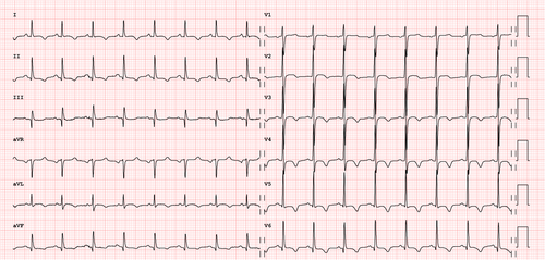
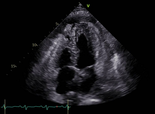
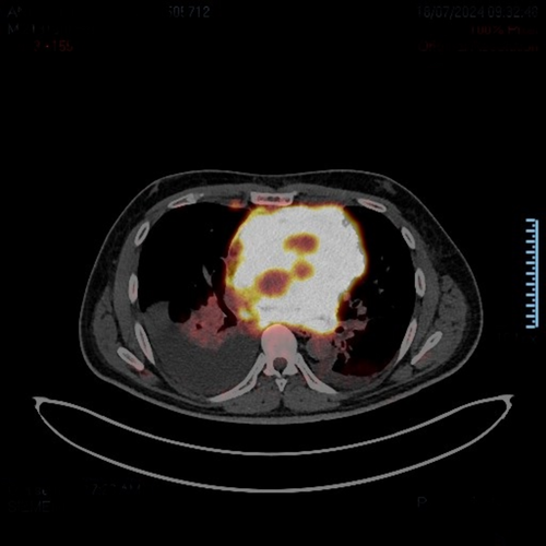
An electrocardiogram revealed inverted T waves in precordial leads from V2 to V6 and in DI, DII, aVL, and aVF (Figure 1). A chest X-ray showed mediastinal enlargement, while echocardiography revealed hyperechogenic areas in the anterior, lateral, and inferior walls of the left ventricle, along with hyperechogenicity of the pericardium and a severe reduction in the left ventricular ejection fraction (LVEF 33%) (Figure 2). Finally, a PET-CT scan was performed, revealing abnormally increased fluorodeoxyglucose (FDG) uptake at the mediastinal level, with extension into the pericardium (Figure 3) The mass biopsy confirmed the diagnosis of primary mediastinal large B-cell lymphoma [2]. The patient was started on chemoimmunotherapy.


The main complications included febrile neutropenia and thrombocytopenia, which required broad-spectrum antibiotic therapy and deep vein thrombosis related to a central venous catheter. The treatment was continued for a total of six chemotherapy cycles.
A PET-CT scan performed after the first two cycles revealed significant dimensional and metabolic mass regression at the mediastinal level, with resolution of the pericardial involvement. Furthermore, echocardiography showed a clear reduction in the hyperechogenic areas of the left ventricle, along with an almost completely recovered LVEF and the ECG improved with a partial normalization of the T waves in precordial leads and in DII and aVF (Figure 4).

3 Discussion
According to literature, the prognosis of B-cell non-Hodgkin's lymphoma with cardiac involvement is generally unfavorable. Chemotherapy is the cornerstone of treatment, leading to prolonged median survival and durable remissions. Radiotherapy is typically reserved for cardiac masses that progress despite chemotherapy due to cardiovascular side effects [3].
The primary, noninvasive approach for morpho-functional assessment is the echocardiogram, which allows for the detection of cardiac involvement and complications. Its main limitation is the small acoustic window it provides.
Multimodal imaging is useful for localization and diagnosis. In this case, echocardiography was sufficient to determine the clear reduction of cardiac infiltration after a single cycle of chemotherapy, and upon completing all six cycles, achieved a complete remission. An echocardiogram performed at the end of the treatment shows a full recovery of the ejection fraction up to 65%.
It is evident that optimal chemotherapic treatment results in rapid improvement of cardiac dysfunction, without the need for additional cardiac pharmacotherapy to treat the myocardal involvement, which in this case is caused by an extrinsic infiltration. Moreover, although ECG is generally not considered sensitive for detecting cardiac mass infiltration, it can reveal conduction abnomarlities due to lymphomatous cell infiltration of the electrical conduction system, like atrial arrythmias and atrioventricular blocks, right bundle block, inverted T waves, low voltage, and ventricular tachiarrhythmia [3].
Author Contributions
Rossana Izzo: conceptualization, investigation, methodology, project administration, writing – original draft. Daniela Campanale: conceptualization, writing – review and editing. Mattia Duchini: conceptualization, data curation, writing – review and editing. Angelo Di Simone: methodology, writing – review and editing. Andrea Menafoglio: methodology, writing – review and editing. Andrea Demarchi: conceptualization, investigation, methodology, project administration, writing – original draft.
Consent
Written informed consent was obtained from the patient to publish this report in accordance with the journal's patient consent policy.
Conflicts of Interest
The authors declare no conflicts of interest.
Open Research
Data Availability Statement
Data sharing is not applicable to this article as no new data were created or analyzed in this study.


