Clinical and radiographic outcomes of maxillary lateral incisors rehabilitation using Morse taper connection extra-narrow implants at 12-month follow-up: A case report
Abstract
Narrow-diameter implants (≤3.5 mm) have been proposed to address the challenge of implant placement in cases of insufficient bone quantity, thin alveolar crest, and small cervical diameter teeth replacement The aim of this study is to report one-year outcomes of extra-narrow implant rehabilitation of maxillary lateral incisors, due to agenesis, in a young adult that presented sites with reduced mesiodistal and buccolingual dimensions. A 26-year-old male patient in need of fixed-implant supported prostheses due to the absence of permanent maxillary lateral incisors and with limited space, was submitted to surgery to receive two 2.9 mm hybrid Morse taper connection implants with hydrophilic surfaces. Immediate loading was applied by means of insertion of provisional prostheses, which were replaced for all-ceramic prostheses 12 months after surgery. The 1 year follow-up showed clinical and radiographic success of extra-narrow implant rehabilitation. Also, both regions presented good evolution of peri-implant esthetics, as assesses using the pink esthetic score, with improvements at 4 months follow-up and reaching high scores 12 months after surgery. Although the prosthetic rehabilitation of maxillary lateral incisors is challenging due to limited space for the insertion of implants, the clinical case suggests that the use of extra-narrow Morse Taper implants with hybrid design and hydrophilic surface is a reliable alternative, presenting good outcomes regarding hard and soft tissue and it is a versatile solution or immediate loading procedure. Further studies are needed to confirm extra-narrow implant predictability.
1 INTRODUCTION
Osseointegrated implants were initially introduced for rehabilitation with multiple or full-arch fixed prostheses and have presented high success rates over the years.1-3 Dental implant placement requires adequate bone volume and horizontal interdental space; therefore, single-tooth restorations have shown to be more difficult due to limited area.4 This is especially common in central and lateral incisors rehabilitation, for which the use of standard diameter devices may lead to the exposure of implant threads, jeopardizing the esthetic results.5
Narrow-diameter implants (≤3.5 mm) have been proposed to address the challenge of implant placement in cases of insufficient bone quantity, thin alveolar crest, and for the replacement of teeth with small cervical diameter.6, 7 Among the advantages of their use, the most significant is the avoidance of bone augmentation procedure which presents some side effects such as edema, pain, discomfort, and risk of nerve injury.8 Moreover, the cost of standard diameter implant placement along with grafting procedures may be inaccessible for some patients. Reduced bleeding and patient morbidity, minimized postoperative discomfort, and lower healing time have also been reported, which may result in patient preference for this treatment option rather than more invasive techniques.4, 9
Although increased crestal bone loss has been suggested to occur after narrow implant placement, especially in the anterior region,10 it has been demonstrated that a distant inadequate implant placement to the adjacent tooth results in loss of bone height, leading to compromised papillae final position6 Thus, by using narrow implants, this distance is increased, which could positively influence papillae formation and lead to better esthetic outcomes.11
This report presents the successful use of extra-narrow implants for maxillary lateral incisor rehabilitation, due to agenesis, in a young adult who presented sites with reduced mesiodistal and buccolingual dimensions, considering 1 year of clinical and radiographic follow-up.
2 MATERIALS AND METHODS
2.1 Case presentation
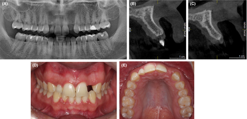
A 26-year-old male patient was addressed to Ilapeo College in need of fixed-implant supported prostheses due to the absence of permanent maxillary lateral incisors, one of these sites with the primary tooth still present. Cone beam computed tomography, periapical X-ray, and photographs were obtained for diagnostic and planning purposes (Figure 1), which confirmed agenesis of lateral incisors (sites 12 and 22, according to FDI tooth number system) and showed that the orofacial bone volume was not enough for conventional implant placements. The patient did not report any health condition but presented gingivitis. Thus, oral hygiene instructions were given with proper brushing technique and flossing before surgery and during the whole follow-up period. Since he did not want to have his teeth worn out for bridge placement, prosthetic rehabilitation with extra-narrow implants was chosen. Written consent was given by the patient.
2.2 Surgical and prosthetic protocol
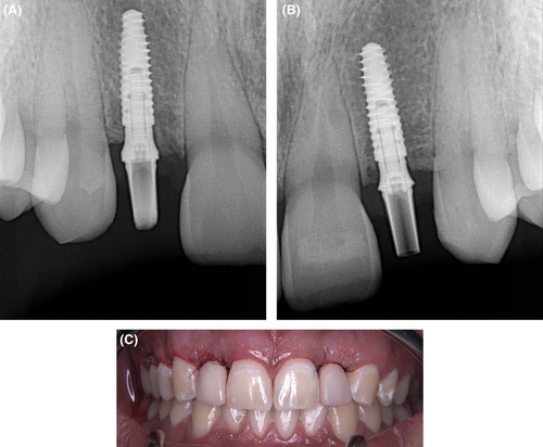
Site preparation was performed as recommended by the manufacturer for bone type II,12 with 500 rpm rotation and adequate irrigation. Two 2.9 × 10 mm hybrid (cylindrical contour on the coronal and conical on the apical part) narrow implants with Morse taper connection (Helix Narrow GM, Neodent,) and hydrophilic surfaces (Acqua, Neodent) were placed at a 2 mm subcrestal bone level, at first using the contra-angle, and finishing with the torque wrench (Figure 2). Final torque of 32 N.cm was obtained for implant 12 and 45 N.cm for implant 22, allowing immediate loading. Thus, 17-degree angled narrow universal abutments (3.3 × 6 × 2.5) were selected and screwed onto the implants to support cement-retained acrylic provisional prostheses (Figure 3). Sutures were placed to close the wounds. Periapical X-rays were taken to check the correct implant position. The patient received post-surgical prescription of antibiotics and analgesics, and oral hygiene instructions were given. Sutures were removed 10 days thereafter.

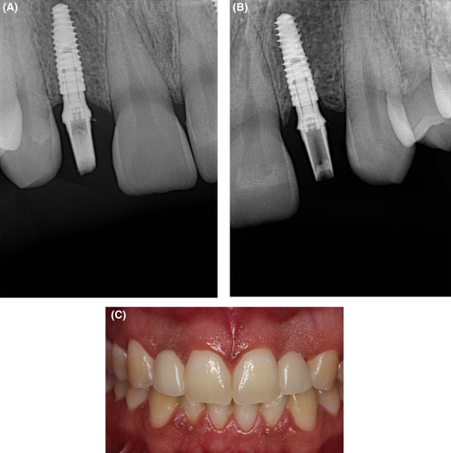
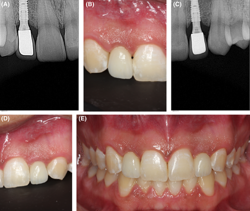
Four months later, clinical and radiographic evaluation revealed adequate peri-implant bone regeneration and satisfactory soft tissue stability (Figure 4). One year after surgery, abutment impression copings were inserted to obtain conventional impressions with addition silicone material (Kulzer,) and well as anatomical impressions of the antagonist arch and bite registration, for the confection of the final prostheses. Then, patient returned for zirconia copings try-in (Figure 5). Finally, after ceramic application, final crowns were cemented over the initially inserted universal abutments (Figure 6). One-year clinical and radiographic implant success could be observed for both implants, with bone level maintenance, absence of peri-implant radiolucency, and no signs of mobility or pain.13

2.3 Soft-tissue evaluation
In order to evaluate peri-implant soft tissue aspect outcomes, the pink esthetic score (PES)14 was applied at 1, 4, and 12 months after surgery. PES is based on the presence of mesial and distal papillae, soft-tissue level and contour, alveolar process profile, and soft-tissue color and texture, as in comparison with a corresponding tooth. For each variable, a 2–1-0 score is given, being 14 the highest score possible, representing ideal peri-implant soft-tissue esthetic aspects.
Both regions presented good evolution of peri-implant esthetics, with improvements already observed at the 4 months follow-up and reaching high scores 12 months after surgery (Table 1). Region 12 presented improvements in the mesial papillae, soft-tissue level, contour, and texture aspects. The other evaluated factors were maintained, and the total score after 12 months was 11. In region 22, the obtained score was even higher, reaching a total of 13, with improvements of all soft-tissue aspects except for alveolar process deficiency, which was observed to be slight at all times.
| Follow-up | Site position | Mesial papilla | Distal papilla | Level of soft tissue | Soft-tissue contour | Alveolar process | Soft-tissue color | Soft-tissue texture | Score |
|---|---|---|---|---|---|---|---|---|---|
| 1 month | 12 | 1 | 1 | 1 | 1 | 1 | 1 | 1 | 8 |
| 22 | 1 | 0 | 1 | 1 | 1 | 1 | 2 | 7 | |
| 4 months | 12 | 2 | 2 | 2 | 1 | 1 | 1 | 2 | 11 |
| 22 | 2 | 0 | 1 | 1 | 1 | 1 | 2 | 8 | |
| 12 months | 12 | 2 | 1 | 2 | 2 | 1 | 1 | 2 | 11 |
| 22 | 2 | 2 | 2 | 2 | 1 | 2 | 2 | 13 |
Note
- At the site position column, numbers are related to FDI classification. At the Score column, numbers are related to total score of pink esthetic evaluation.
3 DISCUSSION
Narrow implants are an excellent alternative for fixed rehabilitation of areas with reduced bone volume, without grafting procedures. Although the literature is not clear about the definition of narrow implants, a systematic review that included implants with diameters smaller than 3.75 mm showed that their survival rates, complications, and bone loss seemed to be no different from those reported for regular diameter implants.15
In this present case, the patient needed to rehabilitate two agenesic maxillary lateral incisors, and since there was reduced buccolingual bone volume as well as mesiodistal space, hybrid morse tapper connection extra-narrow implants (Helix Narrow GM, Neodent,) were chosen to avoid peri-implant fenestration and the need to perform guided bone regeneration procedures.8 Although maxillary bone quality has been reported to be inferior compared to mandibular bone, good primary stability was achieved and immediate loading could be applied, which is an important factor for patients in need of rehabilitation in the esthetic zone.16
Some authors have reported that narrow-diameter implants lead to reduced bone-implant contact area and, consequently, compromised osseointegration.17 Nevertheless, in the present clinical case, successful osseointegration was achieved and maintained at the 12 months clinical and radiographic follow-up, as observed in other studies with long-term follow-up.4, 18
Marginal bone loss, especially after prosthesis placement, has also been suggested to be more expressive in narrow implants.19, 20 In the present study case, although some bone remodeling could be observed, it was not significant (lower than 1.5 mm), and both hybrid Morse taper connection extra-narrow implants were considered successful after one year21
Esthetic considerations are mandatory when planning the insertion of implants in the anterior zone. Regardless of limited space, it must be possible to obtain a natural emergence profile and healthy soft tissues, meeting patients´ expectations.
It has been reported that the ideal distance between implant and adjacent teeth should be of at least 1.5 mm to prevent peri-implant crestal bone remodeling.19 This way, narrow implants are able to fit between natural teeth and limit the interproximal bone resorption that would usually be observed if conventional implants were inserted too close to adjacent teeth.22, 23 Moreover, it has been shown that peri-implant papillae is dependent on the interproximal bone height of the adjacent teeth11.
Studies that evaluated soft-tissue outcomes using narrow implants have reported excellent results, with increasing papilla index scores during the first year, including those with immediate provisionalization.24, 25 In the present case, soft-tissue esthetics improved during the first year, reaching high scores at the 12 months follow-up. Similar results have been found in a previous study, in which 87 narrow implants presented a PES score average of 10.72 ± 2.6 at the 1 year follow-up.23 On the contrary, a mean lower PES score (7.8) has been reported by a different study in which regular-diameter implants were inserted in the anterior area, indicating that better soft-tissue outcomes may be achieved when using narrow implants in this region.22
4 CONCLUSION
Although the prosthetic rehabilitation of maxillary lateral incisors is challenging due to limited space, the clinical case suggests that the use of extra-narrow Morse Taper implants with hybrid design and hydrophilic surface is a reliable alternative, presenting good outcomes regarding hard and soft-tissue outcomes and it is a versatile solution for immediate loading procedures. Further long-term controlled studies with larger samples are needed to confirm extra-narrow implant predictability.
AUTHOR CONTRIBUTIONS
Geninho Thomé involved in conceptualization and investigation. Camila Vianna involved in writing–original draft preparation. Waleska Caldas involved in writing–review and editing. Sergio Bernardes involved in investigation. Carolina Cartelli involved in data curation. Jean Uhlendorf involved in investigation. Larissa Trojan involved in supervision and project administration.
ACKNOWLEDGEMENT
None.
FUNDING INFORMATION
This study was sponsored by Neodent.
CONFLICT OF INTEREST
The authors declare that they work for the company that provided the devices used in this study.
CONSENT
Written informed consent was obtained from the patient to publish this report in accordance with the journal's patient consent policy.
Open Research
DATA AVAILABILITY STATEMENT
The data that supports this study is within the article


