Anal polyp
Abstract
Our case accurately describes a not infrequent finding that is not well understood by endoscopists. Fibroepithelial polyps are benign and should be considered in the differential diagnosis of anorectal polyp.
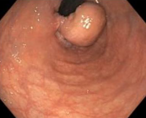
A 75-year-old woman underwent colonoscopy for iron deficiency anemia. Rectal retroflexion revealed a 2.5-cm pedunculated polyp originating from the dentate line [Figure 1]. The polyp was removed completely via transanal excision. What is the etiology?

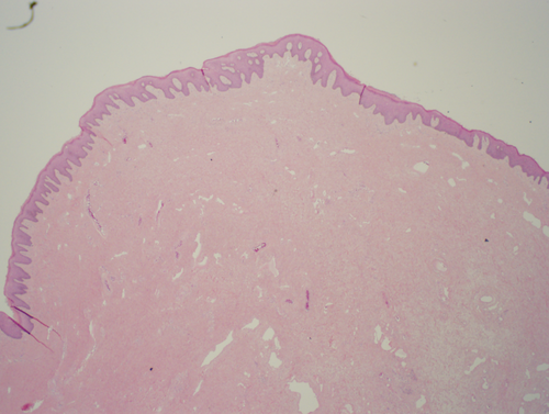
Histopathological examination showed fibrovascular stroma with squamous epithelium suggestive of fibroepithelial anal polyp (FAP) [Figure 2]. FAPs are benign and considered to originate from anal papillae. FAP represents a hypertrophic response to chronic irritation and commonly associated with chronic fissure. They are usually small in size and considered as normal anatomic variation. Large FAPs are rare and should be differentiated from hemorrhoids, adenoma, submucosal tumor, and carcinoma. Large FAPs can cause bleeding, prolapse, and discomfort.1 Endoscopic findings that help to distinguish FAP from adenoma includes their origin from the squamous side of the dentate line and pain with manipulation. Also, biopsy of the lesion always reveals squamous epithelium. Therefore, FAP should be considered in the differential diagnosis of anorectal polyp.

AUTHORS CONTRIBUTIONS
RB, AA, and JA conceptualized and designed the study. RB, US, and AW drafted the article. RB, AA, and US edited the images. RB, AA, JA, AW, and US contributed to the final approval of the article.
ACKNOWLEDGMENT
None.
CONFLICT OF INTEREST
All authors declare that no conflicts of interest or financial relationships exist.
CONSENT
Written informed consent was obtained from the patient to publish this report in accordance with the journal's patient consent policy.
Open Research
DATA AVAILABILITY STATEMENT
Data sharing is not applicable to this article as no new data were created or analyzed in this study.


