A surgical treatment for frank rupture of acute type a small intramural hematoma
Abstract
A 71-year-old woman was admitted to the hospital due to cardiac tamponade. Computed tomography revealed that the diameter and wall thickness of the ascending aorta were 36 and 9 mm, respectively. An emergent ascending aortic replacement was performed uneventfully. The pathological findings indicated frank rupture of intramural hematoma.
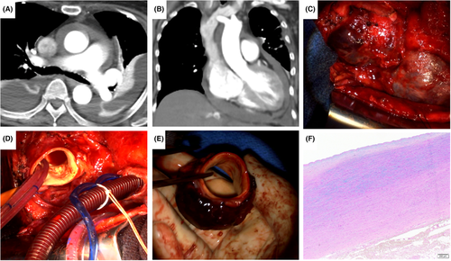
The treatment of acute type A intramural hematoma (IMH) has been controversial. It is reported that initial medical treatment with blood pressure and pain control and repetitive imaging may be a reasonable option, particularly in the absence of aortic dilation (<50 mm) and IMH thickness <11 mm.1 A 71-year-old woman was admitted to the hospital due to cardiac tamponade. Computed tomography revealed that the diameter and wall thickness of the ascending aorta were 36 and 9 mm, respectively, indicating type A intramural hematoma (Figure 1A,B). An emergent ascending aortic replacement was performed, but it resulted in uneventful outcomes (Figure 1C–E). The pathological findings indicated frank rupture of intramural hematoma (Figure 1F). Acute type A small IMH is rare; however, physicians should be aware of this possible complication.

AUTHOR CONTRIBUTIONS
DU involved in conceptualization and investigation and writing the manuscript. KN involved in case presentation section and interpreted the patient data. HN obtained consent for participation and publication from patient's parents and writing the manuscript. YT involved in evaluation of case reports and writing the manuscript. KY involved in reviews/edits of case report write-up and appropriate source citation. All authors read and approved the final manuscript.
ACKNOWLEDGMENT
None.
CONFLICT OF INTEREST
The authors declare no conflict of interest.
CONSENT
Written informed consent was obtained from the patient to publish this report in accordance with the journal's patient consent policy.
Open Research
DATA AVAILABILITY STATEMENT
All relevant data are within the manuscript and its Supporting Information files.


