An epidermal cyst in the areola
Abstract
Although epidermal cysts are common lesions of the scalp, face neck, and trunk, these cysts are rarely found in the areola. Doctors should think of epidermal cyst as one of differential diagnoses of an areolar lesions.
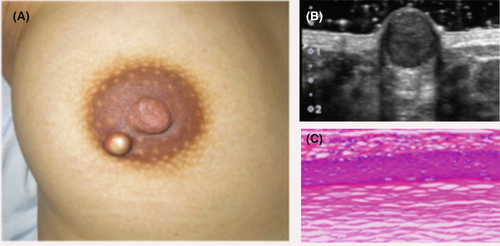
A 28-year-old woman presented to the outpatient breast clinic with a lump in the areola of her right breast. The lesion was first noticed 2 months earlier and had gradually increased in size. On examination, the tumor was approximately 1 cm in diameter, well-defined, and dome-shaped, while the overlying skin was glossy and whitish (Figure 1A). Ultrasound showed a well-circumscribed hypoechoic solid mass (Figure 1B). The tumor was excised under local anesthesia. Histological examination demonstrated that the resected lesion was a cyst lined by mature stratified squamous epithelium and filled with laminated layers of keratin. These findings were consistent with a diagnosis of epidermal cyst (Figure 1C). Although epidermal cysts are usually situated in the scalp, neck, and trunk, while they are rarely found in the nipple-areolar complex.1 Doctors should think of epidermal cyst as one of differential diagnoses of an areolar lesion. Given complications in the future and patient’s physical and psychological discomfort, surgical removal is the most appropriate management option.2

AUTHOR CONTRIBUTION
The author has been responsible for data collection, analysis, and presentation.
ACKNOWLEDGMENT
The author would like to thank the patient for giving consent.
CONFLICT OF INTEREST
Nothing to declare.
ETHICAL APPROVAL
This article does not contain any studies with human participants or animal subjects.
CONSENT
Written informed consent was obtained from the patient to publish this report in accordance with the Journal's patient consent policy.
Open Research
DATA AVAILABILITY STATEMENT
All data relevant to the study are included in the article.


