Atypical lipomatous tumor of the oropharynx: A case report
Abstract
Atypical lipomatous tumor or well-differentiated liposarcoma is a rare condition in the laryngopharynx. We present a case of a 63 year-old male with progressive dysphagia who later manifested with a mass that regurgitates outside the oral cavity. Surgical removal was done via a transoral approach, followed by adjuvant radiotherapy.
1 INTRODUCTION
Atypical lipomatous tumor, also known as well-differentiated liposarcoma is a subtype of liposarcoma based on the WHO classification of soft tissue tumors. It is one of the most common soft tissue sarcomas in adults, but is a rare pathology in the laryngopharynx with mostly case series or reports available in literature. Symptoms of dysphagia, globus sensation, and regurgitation of a fleshy mass out of the mouth may all be present, similar to the presentation of the condition called fibrovascular polyp. However, further evaluation with immunohistochemistry and molecular studies will help elucidate the diagnosis of an atypical lipomatous tumor.
2 CASE REPORT
A 63-year-old male, previous smoker, came to our institution with a 1 year and 7 months history of progressive dysphagia. Initially, patient experienced dysphagia to solids. No consult was done until after several months, when he experienced intermittent dyspnea. He was given bronchodilators which afforded relief of dyspnea. However, patient now reported globus sensation and bloatedness. He sought consult with another physician and was prescribed with Omeprazole for possible reflux for 4 months.
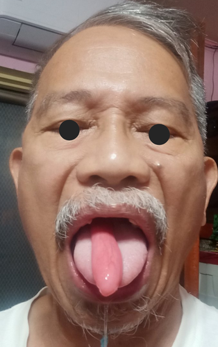
In the interim, patient experienced an episode of sudden protrusion of a fleshy pedunculated mass outside his oral cavity after coughing (Figure 1). There were two more instances when this occurred and what patient did was to swallow successively until the mass lodged back inside. His dysphagia progressed as he reported occasional difficulty of swallowing liquids. He also had significant weight loss of about 9 kg within 2 months. Patient again consulted with another physician and was advised to undergo esophagogastroduodenoscopy (EGD), which revealed a smooth, pedunculated mass originating from the hypopharynx and extending toward the esophagus. Patient was then referred to our institution for excision of the mass.

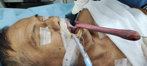
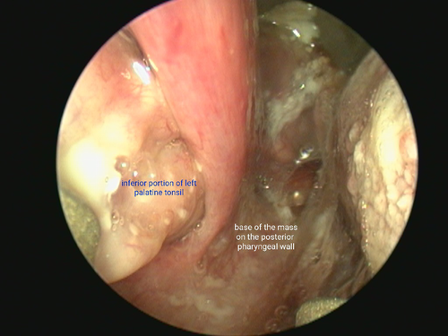
When the patient was referred for surgical management, a possible fibrovascular polyp was considered. Repeat EGD was carried out to confirm its location. The base of the mass was noted in the oropharynx about 12 cm from the incisors, which insinuated through the pyriform sinus. Upon advancing the scope further into the esophagus, the distal end was found at 35 cm from the incisors. The distal end was grasped with a rat tooth forceps and pulled out from the esophagus toward the oral cavity, and tied using a nonabsorbable suture for traction during excision (Figure 2). Exposure of the base of the mass, which originated from the posterior pharyngeal wall adjacent to the inferior portion of the left palatine tonsil was done (Figure 3). Excision via transoral approach was performed using KTP laser and completed using bovie cautery. Specimen obtained was a smooth mass, about 15 cm in length, with a 1 cm diameter on its proximal end and 3.3 cm diameter on its distal end (Figure 4). Patient was discharged the following day without complications and with resolution of previous symptoms.


Final histopathology later showed a spindle cell neoplasm with tumor present at the proximal margin of resection. Immunohistochemistry studies were suggested for further evaluation with the following results: Mouse double minute 2 (MDM2) positive (Figure 5), S100 positive, CD34 negative, Smooth muscle actin (SMA) negative and Desmin negative. Findings were suggestive of an atypical lipomatous tumor/well-differentiated liposarcoma. Additionally, a Fluorescence In Situ Hybridization (FISH) study for MDM2 gene amplification was done which confirmed the diagnosis. Following the American Joint Committee on Cancer (AJCC) 8th edition staging for soft tissue sarcoma of the head and neck, a diagnosis of atypical lipomatous tumor/well-differentiated liposarcoma of the oropharynx, Stage IB (T3N0M0G1) was given. Adjuvant radiation therapy was delivered via IMRT to a dose of 5000 cGy with a boost to 7000 cGy to the tumor bed due to the presence of a positive pathologic margin. This regimen was consistent with NCCN 2.2022 Soft Tissue Sarcoma Guidelines. Patient is on regular follow-up, and currently with no evidence of disease at 6 months postsurgery.

3 DISCUSSION
Atypical lipomatous tumor/well-differentiated liposarcoma is an extremely rare malignancy in areas of the oropharynx, larynx, or hypopharynx. This pathology usually occurs in the extremities, trunk, and retroperitoneum. Few publications for this type of tumor in the laryngopharynx are available, and most were located in the hypopharynx.1-3 Patients commonly present with symptoms of dysphagia, globus sensation, episodes of intermittent dyspnea, or choking sensation, with an unusual regurgitation of a smooth pedunculated mass. In few cases, patient may even experience asphyxiation prompting an emergency intervention. These symptoms are very similar to a fibrovascular polyp,3 which was initially considered in this patient. Fibrovascular polyp is also a rare entity in the upper aerodigestive tract, but is a benign condition.
Diagnostics for atypical lipomatous tumor/well-differentiated liposarcoma include a modified barium swallow and flexible fiberoptic endoscopy to be able to locate the origin of the mass. An EGD is also done for further evaluation, as the pedunculated mass usually extends toward the esophagus. Computed tomography (CT) and magnetic resonance imaging (MRI) have also been reported in evaluation of these cases. However, definitive diagnosis can only be made on histopathology and confirmed by immunohistochemistry and molecular studies. Morphologic examination with attention to cytologic atypia, as well as fluorescence in situ hybridization for MDM2 amplification, will help confirm a diagnosis of atypical lipomatous tumor/well-differentiated liposarcoma.4 In this patient, the presence of atypical, hyperchromatic spindle cells interspersed with adipocytes of varying sizes raised suspicion of a possible adipocytic neoplasm. Immunohistochemistry results showed positivity for MDM2 gene, and FISH study for MDM2 gene amplification verified it was indeed an atypical lipomatous tumor.
Since a fibrovascular polyp was initially considered in this case, resection of the mass from its base was carried out. However, further studies showed it is otherwise benign. Surgical wide resection is the recommended treatment for patients with atypical lipomatous tumor/well-differentiated liposarcoma, a low grade liposarcoma.3 Due to positive margins noted on final histopathology, either a re-resection or adjuvant radiotherapy may be considered, based on the NCCN guidelines.5 Clear margins are important to minimize risk of recurrence, but may not always be possible due to anatomical constraints.3 Re-resection in case of recurrence due to positive margin in the area of the oropharynx maybe unduly morbid, hence we opted for adjuvant radiotherapy due to risk of local recurrence.
4 CONCLUSION
Atypical lipomatous tumor/well-differentiated liposarcoma of the oropharynx is extremely rare but can present with nonspecific symptoms such as dysphagia. The presence of a mass warrants surgical excision; however, gross examination and endoscopic findings cannot easily differentiate this condition from other benign pathologies of the laryngopharynx. Immunohistochemistry and molecular studies are therefore needed for definitive diagnosis to determine the optimal management for these cases.
AUTHOR CONTRIBUTION
APCD prepared the manuscript with contribution from all coauthors.
ACKNOWLEDGMENTS
The authors acknowledge Dr. Abigail Sarmiento-Salcedo for helping with the documentation of this patient.
CONFLICT OF INTEREST
The authors have no conflicts of interest to disclose.
ETHICAL APPROVAL
The manuscript related to human use has been complied with all the relevant national regulations, institutional policies, and in accordance the tenets of the Helsinki Declaration, and has been approved by the authors' institutional review board, University of the Philippines Manila Research Ethics Board (UPMREB) and has been given a certificate of exemption (RGAO-2022-0075).
CONSENT
Written informed consent was obtained from the patient to publish this report in accordance with the journal's patient consent policy.
Open Research
DATA AVAILABILITY STATEMENT
The data that support the findings of this study are available from the corresponding author upon reasonable request.


