Pseudoaneurysm of the proximal superficial femoral artery following blunt trauma in a footballer: A rare case report
Abstract
Pseudoaneurysm of superficial femoral artery without any femoral fracture following blunt trauma is a rare clinical entity. Most cases of pseudoaneurysm of superficial femoral artery present to the hospital following penetrating injury, femoral fracture, and surgical procedures. Failure of management of pain despite taking analgesics should lead to suspicion of pseudoaneurysm.
1 INTRODUCTION
Pseudoaneurysm is a hematoma not contained by vessel walls, but rather contained by a thin fibrous capsule.1 It is also known as false aneurysm. Pseudoaneurysm of superficial femoral artery without penetrating injury or femoral bone fracture is a rare clinical presentation.2 It is very important to think of pseudoaneurysm of femoral artery in case of blunt trauma when the pain has not subsided after taking Non-steroidal anti-inflammatory drugs and opioid analgesics. Here, we report a case of a 20 years footballer who presented to our hospital with complaints of pain and swelling in right thigh for 2 weeks.
2 CASE REPORT
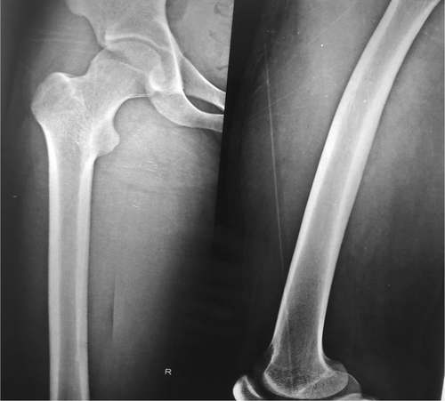
A 20-year-old footballer presented to the out-patient department of cardiothoracic and vascular surgery of Metrocity Hospital with complaints of pain and swelling in the right thigh for 2 weeks. Pain was acute in onset following the trauma, non-radiating, pricking type, continuous in nature, not responding to non-steroidal anti-inflammatory and opioid analgesics. Initially, the severity of pain was 4/10 on the visual analogue scale, but since 2 days it has increased to 7/10. The patient was seen initially in the local hospital where he underwent X-ray, right thigh. X-ray of the right thigh did not reveal any fractures (Figure 1).
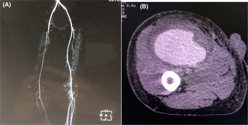
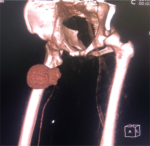
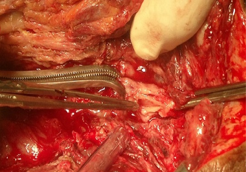
So, he was prescribed tablet ibuprofen 400 mg thrice a day. However, despite taking ibuprofen for 7 days, his pain did not subside. So, he was prescribed oral tramadol 100 mg thrice a day. Even after taking tramadol for 5 days, his pain did not subside. So, he was referred to Manipal teaching hospital, where he underwent ultrasound of the swelling on the right thigh and Doppler study of the arterial and venous system of the right lower limb. USG revealed a rounded heterogeneous area measuring 8.9 × 6.2 cm with a predominant hypoechoic component in the right anteromedial thigh in proximity to the right superficial femoral artery with a Ying yang sign on color flow and to and fro pattern seen with pulsed Doppler due to turbulent flow suggestive of right superficial femoral artery aneurysm. So, he was referred to our hospital as we are the only hospital providing vascular facility in Pokhara, Nepal. The patient underwent CT angiography of the lower limb where there was 8.8 × 6.3 cm lesion at the upper third of the right thigh anteriorly displacing the muscles of the anterior compartment (Figure 2A). Post-contrast study revealed homogenous enhancement surrounded by hyperdense area suggestive of hematoma (Figure 2B). Posteriorly, that lesion was communicating with the superficial femoral artery, suggestive of a right superficial femoral artery aneurysm (Figure 3). So, he was diagnosed as a case of pseudoaneurysm of right superficial femoral artery. The patient was then planned for surgery. Then, SFA was explored, which revealed there was about 10 ml of sero-sanguinous collection and a 2 cm contused segment of vessel about 11 cm proximal to the knee joint. The pseudoaneurysm was resected (Figure 4), fogartisation of both proximal and distal ends were done followed by anastomosis of proximal and distal ends. Medial and lateral fasciotomy was done on the right leg at the end.
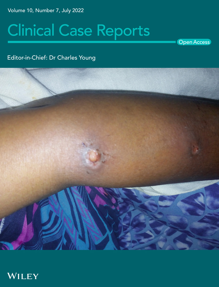
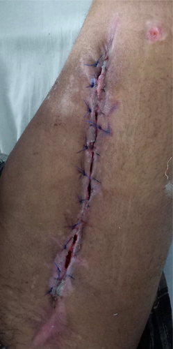
Post operatively, heparin was started after 24 h of surgery in the patient. The limb was warm with normal capillary refill time. However, there was a surgical site infection after a week. Wound dressing was done regularly, which was later followed by secondary suturing on the 15th postoperative day.(e4 Figure 5) After 30 days, Doppler study of the arterial system in the lower limb revealed normal flow in common femoral, superficial femoral, deep femoral, popliteal, anterior tibial, and posterior tibial artery. Follow-up of the patient at 3 months of follow-up was normal with no abnormality on Doppler study.
3 DISCUSSION
The femoral artery supplies blood solely to the lower extremity. The femoral artery courses in the femoral triangle of the thigh before dividing into superficial and profunda femoris.3 Superficial femoral artery further divides into five branches. They are the superficial circumflex iliac, superficial epigastric, superficial external pudendal, deep external pudendal, and descending genicular artery. The profunda femoris terminates in the thigh. It is important to know the anatomy of the femoral artery for early diagnosis of the injury to the femoral artery.
Femoral artery injury is a rare clinical scenario. It is commonly seen in association with open injuries, penetrating injury, and femoral bone fracture. It is very rare to see isolated femoral artery injury without any injury to the bone. In 1968, motor scooter handlebar syndrome was described where there is injury to the common femoral artery without pelvic or femoral fracture.4 This syndrome was described as a result of compression of the artery by the inguinal ligament.
In the past, isolated injury to the superficial femoral artery has rarely been described. Our case is one of the rare cases where clinician often fail to make a suspicion. The patient was suspected of soft tissue injury and prescribed tablet ibuprofen initially that was later followed by Cap Tramadol. However, persistence of pain even after taking analgesics made us think of vascular injury.
Close proximity of the superficial femoral artery after passing through Hunter's canal and its superficial location makes it vulnerable for injury following blunt trauma.5
Blaise and Pape also reported a similar case, but it was an isolated pseudoaneurysm of the deep femoral artery.2 The pseudoaneurysm was excised in their case as we did in our case, but in the superficial femoral artery.
Apart from these, there are other techniques where radiological intervention has successfully treated pseudoaneurysm. In the study done by Saptarishi et al.,1 they have treated the pseudoaneurysm by the endovascular coil embolization. However, they treated the case of pseudoaneurysm of deep femoral artery.
So, it is important for clinicians to suspect vascular injury in case of blunt trauma where there is no injury to the bones. A clinician must be vigilant to identify such injury early enough to prevent the complications.
4 CONCLUSION
Isolated pseudoaneurysm of femoral artery following blunt trauma is a rare pathology. In a case of blunt trauma, it is necessary to suspect pseudoaneurysm where there is no bone injury and the pain is not responding despite taking painkillers, as in our case.
AUTHOR CONTRIBUTIONS
AP, BJK, SB, KGS, and GRP conceptualized the study and preparation of the manuscript. AP and BJK involved in design of the manuscript. SB and GRP involved in literature review.
ACKNOWLEDGMENT
None.
CONFLICT OF INTEREST
None.
CONSENT
Written informed consent was obtained from the patient for publication of this case report and any accompanying images.
Open Research
DATA AVAILABILITY STATEMENT
The first author and corresponding author will provide necessary information on request from the editor-in-chief.


