Case of multiple furuncular myiasis in Northeast Ethiopia
Abstract
Human myiasis is a neglected disease caused by parasitic infestation of the skin, cavities, and other body parts by larvae (maggots) of a wide variety of dipteran flies. Here, we present a case of multiple furuncular myiasis caused by Cordylobia anthropophaga in a 61-year-old woman from Northeast Ethiopia.
1 INTRODUCTION
Human myiasis is a neglected disease caused by parasitic infestation of the skin, wound, cavities, and other body parts by larvae (maggots) of a wide variety of dipteran flies. Myiasis has a global sporadic distribution. However, it is more frequently reported from tropical areas in Africa. Numerous types of myiasis could occur in humans, and it is mainly determined by the presence and geographic distribution of different fly species.1 Based on parasite–host interaction, myiasis can be classified broadly as obligatory, facultative, and accidental myiasis. It can also classify as cutaneous, nasopharyngeal, intestinal, and urogenital myiasis on the basis of body parts affected.1, 2 The dipterous larvae species causing obligatory myiasis requires a living host tissue for its survival.3
Cutaneous myiasis is the most common forms of human myiasis which occurs after the penetration of dipteran larvae into the skin. Furuncular, migratory, and wound myiasis are the three clinical forms of cutaneous myiasis. However, furuncular myiasis is the major type that usually manifested with papular and nodular skin lesions.4 Cordylobia anthropophaga and Dermatobia hominis are the primary fly species identified for the distribution of furuncular myiasis in Africa and Latin America, respectively.5, 6 In general, myiasis is underestimated and unfamiliar problem for most of the healthcare providers in Ethiopia. Thus, we present a case of multiple furuncular myiasis in a 61-year-old woman.
2 CASE PRESENTATION
A 61-year-old woman from Kombolcha town in Northeast Ethiopia was presented to Kombolcha 03 health center with a chief complain of swelling over the left arm, abdomen, and left thigh. She had also pain, discomfort, and itching sensation over the swelled areas. She had no history of any skin lesion in the past and no family history of boil or carbuncle. She had no history of hypertension, diabetes, heart failure, surgery, trauma, and bite. Moreover, she lives in a single room house that has no private latrine and garbage cans were available near her house.
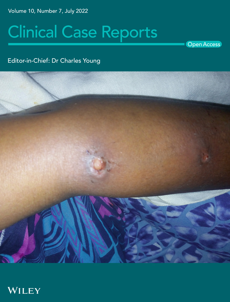
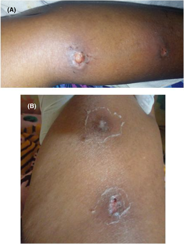
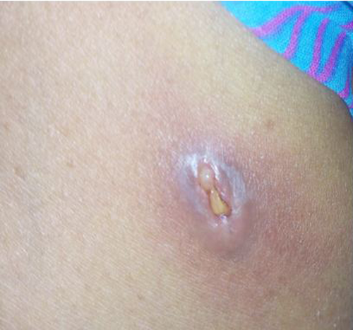
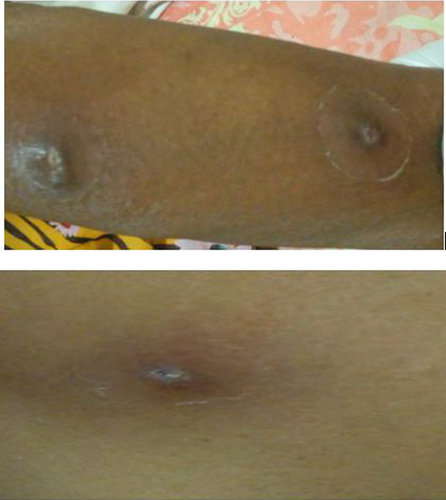
The skin examination revealed tender, erythematous, soft and edematous masses with open skin lesions. Two of the edematous lesions were detected in the left arm with no bleeding and pus discharge. However, the posterior ends of the larvae were detectable protruding from the centre of the lesions at the proximal end of the lower left arm (Figure 1A) and on the left thigh (Figure 1B). The other lesion with visible larva was detected in the abdomen (Figure 2).
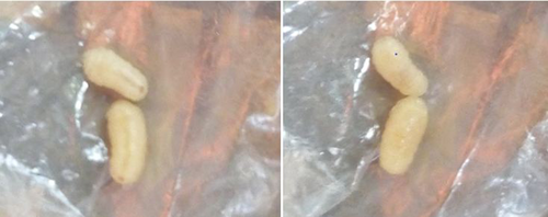
The larvae were removed manually at the proximal end of the lower left arm and abdomen (Figure 3). A diagnosis of multiple furuncular myiasis was done based on the clinical manifestations and the presence of larvae. As it was not possible to send the specimens to an entomologist, we used taxonomic keys from the literature to identify the larvae to those of C. anthropophaga.7, 8 Wound care using hydrogen peroxide and normal saline was provided for 7 days and aseptic technique was implemented throughout the procedure. After the removal of the larvae and daily wound care, the patient was relatively recovered with the formation of circular remnant scars and crust at the center (Figure 4).
3 DISCUSSION
Human myiasis has been reported frequently from tropical countries, particularly in Africa and Latin America.5, 9 It can occur at any age of the life and cutaneous myiasis is the prominent type of human myiasis.4 Furuncular myiasis, which is the main form of cutaneous myiasis can be diagnosed clinically. However, a routine dermoscopy is highly recommended for precise diagnosis of furuncular myiasis and sometimes ultrasound can be used.10 In our case, the posterior ends of the larvae were detectable protruding from the center of the lesions and C. anthropophaga was identified using literature taxonomical keys. In Africa, C. anthropophaga and C. rodhaini are the main etiologic agents causing furuncular myiasis. However, D. hominis have been reported mainly in America.9
The skin lesions of furuncular myiasis could be localized or multiple. This patient was presented with multiple furuncular lesions in the arm, thigh, and abdomen. The complete removal of larvae from the skin and prevention of secondary infection are the goals of treatment in furuncular myiasis. In addition to the mechanical removal of maggots out of the skin, suffocating and toxic agents such as turpentine oil, petroleum jelly, and ivermectin can be used as required.11 Surgical removal of maggots may be needed in rare cases. In this patient, the larvae were removed manually.
Patients with furuncular myiasis could have associated symptoms of pruritus, pain, skin redness, and sense of crawling beneath the skin.12, 13 In this patient, swelling, pain, and skin redness were the predominant clinical presentations. Papules and nodules are the main skin lesions observed in furuncular myiasis1 and in our case, the papular type was identified. Furuncular myiasis is usually self-limiting, and the lesions can be healed after the removal of larvae. However, bacterial super infection may complicate and prolonged the duration of the lesions occasionally.14, 15 The limitation of this case report is that the species causing the myiasis was not identified on molecular or morphological bases.
4 CONCLUSIONS
Careful examination of all type of skin lesions and high index of suspicion are essential to diagnose multiple furuncular myiasis in clinical areas. Moreover, health professionals in Ethiopia shall be aware of furuncular myiasis to identify and treat similar cases as early as possible.
AUTHOR CONTRIBUTIONS
AD and AK contributed to the collection of data and the management of the patient. AD and SG wrote the initial draft of manuscript. AD, SG, and TD revised and prepared the final version of the manuscript. All authors have read and approved the final manuscript and agree to take full responsibility for the integrity and accuracy of the work.
ACKNOWLEDGMENT
None.
CONFLICT OF INTEREST
None.
CONSENT
Written informed consent was obtained from the patient to publish this report in accordance with the journal's patient consent policy.
Open Research
DATA AVAILABILITY STATEMENT
The data are available from the correspondent author and can be obtained upon request.


