Synovial hemangioma: A rare cause of chronic knee pain
Abstract
Synovial hemangioma is a rare benign vascular tumor responsible for chronic knee pain and swelling. Given its non-specific symptoms, synovial hemangioma is often misdiagnosed. We report a case of synovial hemangioma of bilateral knee joints occurring in a young military man.
1 INTRODUCTION
The knee is a location for many tumors, affecting commonly bones and rarely synovial membrane and soft tissues.1 Several tumors and tumor-like proliferations can occur in the synovial membrane, such as giant cell tumor of the tendon sheath (GCTTS), pigmented villonodular synovitis (PVNS), localized nodular synovitis (LNS), synovial chondromatosis, lipoma arborescens, and synovial hemangioma.
The intra-articular synovial hemangioma is a rare benign vascular tumor of the synovial membrane.2 It can be localized or multifocal in the same joint. It usually affects children or young adults.3, 4 It can be responsible for chronic knee pain and swelling. Since its clinical manifestations are non-specific, synovial hemangioma is often misdiagnosed.
We report a case of synovial hemangioma of bilateral knee joints occurring in a young military man. We emphasize clinical, radiological, and management of this disease.
2 CASE PRESENTATION
A 26-year-old man presented to our department with a four-month history of progressive pain and swelling of the right knee.
This pain was responsible for activity limitation, discomfort, and motion impairment.
There was no history of trauma, fever, or loss of weight and appetite.
He was a military soldier.
Physical examination showed swelling over the right popliteal fossa region. There was no redness or warmth. The visual analogue scale (VAS) was 7 out of 10. He also had a restricted right knee flexion (100°) and a loss of full extension (−5°). There was no joint effusion nor signs of meniscus or ligament injury.
Complete blood cell count, inflammatory biomarkers, and coagulation parameters were within normal ranges.
Knee radiographs did not demonstrate bone erosions or femorotibial and femoropatellar joint space narrowing. The pelvic radiograph was normal.
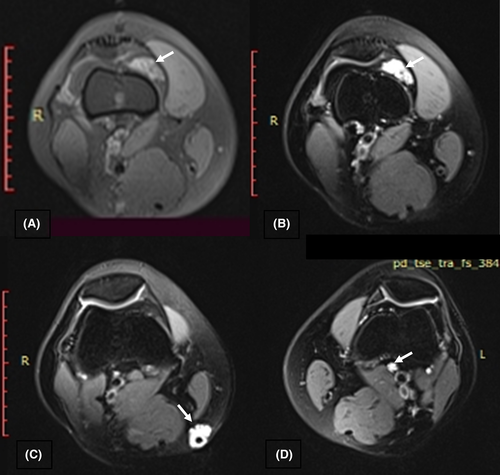
Magnetic resonance imaging (MRI) showed intra-articular lesions affecting both knee joints, appearing like a cluster of grapes (Figure 1). These lesions had an intermediate signal intensity on T1-weighted images and high-signal intensity on T2-weighted images. A diffuse enhancement was seen after Gadolinium administration.

There were no bone erosions or extensions into periarticular soft tissues.
The diagnosis of a synovial hemangioma was made based on the clinical and radiological findings.
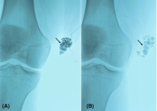
The patients underwent percutaneous sclerotherapy for the synovial hemangioma of the right knee (Figure 2). No complications were noted during and after this treatment. After three months of follow-up, the patient reported alleviation of signs and symptoms. The VAS fells from 7 to 2 out of 10, and he recovered a normal knee range of motion degrees of flexion and extension (130°/0°).

Written informed consent for the case to be published was obtained from the patient.
3 DISCUSSION
A hemangioma is a tumor-like mass resulting from the proliferation of blood vessels. It commonly affects capillaries and veins. It can involve several sites such as the liver, lungs, the digestive tract, the brain, and the spinal cord. The skin and subcutaneous tissue are the most affected sites.
Hemangioma of the synovial membrane is scarce, concerning less than 1% of all hemangioma.5 It mainly involves the knee joint.6
A literature search in MEDLINE for English-language sources yielded 107 articles reporting cases of synovial hemangioma which including 65 cases of knee's location. Only 33 cases were published during the last two decades.
Synovial hemangioma of the knee is often unilateral. We here reported a case of bilateral knee synovial hemangioma.
Synovial hemangioma may also affect the wrist, the elbow, the ankle, and rarely hip joints.7, 8 It commonly appears earlier in young patients, but it can have a delayed onset.9, 10
As in our case, patients may present with joint pain, swelling, and restricted joint motion. Some cases of recurrent joint effusion were reported.11, 12 Symptoms are often non-specific, and the delay to diagnosis can be unacceptably prolonged.
Several diagnoses should be considered in young patients with progressive knee pain and swelling. These symptoms may be due to synovial disorders such as PVNS, synovial chondromatosis, and chronic arthritis; tumor-like lesions such as fibroma of the tendon sheath and synovial lipoma; and malignant tumors such as synovial sarcoma or chondrosarcoma, and synovial metastases.13
Chronic arthritis, synovial sarcoma or chondrosarcoma, and synovial metastases are responsible for inflammatory pain, while synovial hemangioma induces mechanical pain.
Synovial hemangioma has no specific features on radiograph and ultrasonography. At the early stage, radiographs can be without abnormalities. In later diagnosed cases, they may show soft tissue swelling or mass,14 phleboliths, narrowing joint space, and rarely bony erosions.10, 15 Radiographs may also reveal hemophilia-like arthropathy.16 Recurrent hemarthrosis and intra-articular accumulation of mononuclear cells due to hemangioma lead to cartilage destruction and osteoarthritis.17 Doppler ultrasound may reveal a homogenous hypoechoic lesion with internal vascularity.14 Angiography can be indicated in patients with cutaneous hemangiomas or abnormal varicosity. It can reveal feeder's vessels and allow embolization.4
The MRI is the gold standard for the diagnosis of synovial hemangioma. It appears as an intra-articular lobulated mass with an intermediate or isointense signal on T1-weighted images, hyperintensity on T2-weighted images, and intense enhancement after gadolinium administration. The MRI allows assessing the tumor size, its localization, and its extension to soft tissue,1, 15 and it is helpful to exclude differential diagnoses.18
Histological findings of synovial hemangioma provide better diagnostic accuracy. It can specify the predominant type of proliferation: cavernous, capillary, arteriovenous, or venous.1 It can show dilated vascular clusters incorporated in a fibro-myxoid stroma and covered by the synovial membrane.9, 10
However, histological confirmation is not always necessary in patients with typical radiological features of synovial hemangioma.
The treatment of synovial hemangioma is not codified. It may include sclerotherapy,4 selective embolization, arthroscopic excision,19 arthroscopic laser ablation for small-sized tumors, and open excision for large-sized ones.20 After this treatment, pain disappearance and joint motion improvement had been reported.21, 22 However, recurrence is possible in patients with diffuse synovial hemangioma, highlighting the necessity of a long-term follow-up in these patients.
Untreated, synovial hemangioma could be responsible for recurrent hemarthrosis, hemophilia-like arthropathy, and osteoarthritis. Large-sized tumors can progressively infiltrate adjacent muscles and bones, leading to juxta-articular damages.
4 CONCLUSION
Despite its rarity, the diagnosis of synovial hemangioma should be considered in young patients with painful joint associated with swelling. Because of possible functional disability and significant morbidity, physicians should be aware of this disease to make the diagnosis early and to avoid structural damage.
We highlight the importance of MRI features in making the diagnosis of synovial knee hemangioma. Ultrasonography can be also helpful in some cases. Sclerotherapy can be the therapeutic choice for this tumor-like lesion.
AUTHOR CONTRIBUTIONS
Maroua Slouma has substantively revised the work. Emna Hannech has drafted the work. Amjed Msolli and Rim Dhahri have made substantial contributions to the literature research. Imen Gharsallah, Sami Kouki, and Leila Metoui have made substantial contributions to the design of the work. Bassem Louzir has made substantial contributions to the conception of the work.
ACKNOWLEDGMENTS
we would like to thank Dr. Noureddine LITAIEM for language editing.
CONFLICT OF INTEREST
None.
ETHICAL APPROVAL
This manuscript was approved by the ethical committee.
CONSENT
Written informed consent for the case to be published was obtained from the patient.
Open Research
DATA AVAILABILITY STATEMENT
The datasets used during the current study are available from the corresponding author on reasonable request.


