De novo recurrent spontaneous coronary artery dissection in nonatherosclerotic elderly woman: A case report
Abstract
Spontaneous coronary artery dissection (SCAD) is a rare disease which causes acute myocardial infarction (AMI). In this case, we show the recurrence of SCAD and pathological findings of an intimal tear that lead to SCAD of the proximal left anterior descending branch which could contribute to the onset of AMI.
1 INTRODUCTION
Spontaneous coronary artery dissection (SCAD) is an important disease causing acute coronary syndrome (ACS) and is defined as a coronary artery dissection that is not traumatic, iatrogenic, and not with atherosclerosis.1 Generally, SCAD appears to have a particular predominance in young women or people with less conventional atherosclerotic risk factors.2 In pathology, the tunica media is separated from the tunica intima by intramural hemorrhage (IMH), leading to compression of the true lumen. IMH is thought to occur by two mechanisms. The first theory proposes that a disruption of the tunica intima is the initial change, and the second one proposes that a rupture of a single vasa vasorum, which is within the vessel wall and provides the vessel with blood, is the initial change.2, 3 A single vasa vasorum in atherosclerotic coronary arteries has a different character from that of the normal coronary artery and may be fragile and prone to rupture.4 In this report, we detect important pathological findings suggestive of an intimal tear and support the first proposed mechanism of SCAD onset.
2 CASE HISTORY
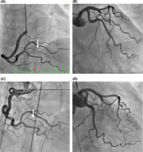
A 57-year-old woman, who had a history of SCAD in the distal right coronary artery (RCA) and continued undergoing conservative therapy, was admitted to our hospital. Three years ago, she visited our hospital for typical chest pain. The electrocardiogram revealed ST-segment elevation in the inferior leads (Figure 1A). The coronary angiography (CAG) showed 90% luminal stenosis in the distal RCA (Figure 2A) and no significant left coronary artery stenosis (Figure 2B). She had no history of diabetes mellitus, dyslipidemia, autoimmune disease, vasculitis, and smoking habit. She also had no family history of connective tissue diseases, such as Marfan's syndrome and Ehlers-Danlos syndrome. After 7 days of observational admission, she continued to visit the outpatient department and was prescribed a calcium channel blocker.


Surprisingly, she presented with cardiopulmonary arrest (CPA) and return of spontaneous circulation (ROSC) in the ambulance and was brought to our emergency department. After ROSC, her systolic blood pressure was 120 mm Hg and her pulse was 80 bpm (sinus rhythm); however, her level of consciousness decreased (E1V1M1). She displayed agonal gasp, and we performed intubation with an artificial ventilator. The electrocardiogram showed tachycardia and ST-segment elevation in the lead I and aVL (Figure 1B). A chest X-ray showed moderate cardiomegaly and pulmonary congestion. The laboratory data showed that AST 521 IU/L, ALT 695 IU/L, LDH 1546 IU/L, Cr 1.01 mg/dL, creatinine kinase-muscle/brain was elevated to 56 IU/L, and high-sensitivity troponin I was 1551 pg/mL. The echocardiogram revealed pericardial fluid and reduced systolic function, and the left ventricular ejection fraction was 40%. In the angiography room, she showed low blood pressure and bradycardia, so we administered noradrenaline, adrenaline, and atropine. However, her hemodynamics collapsed, so we introduced percutaneous cardiopulmonary support (PCPS) and intra-aortic balloon pumping (IABP). Emergent CAG revealed TIMI3 flow (Figure 2C,D). The distal RCA, which had past history of focal SCAD 3 years ago, showed good flow to the distal coronary artery (Figure 2C, arrow). From CAG results, we administered a conservative treatment. Despite our efforts, she suffered a cardiac arrest and unfortunately died on the same day.
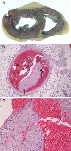
Her postmortem examination revealed extensive acute myocardial infarction (AMI) involving a part of the left ventricular free wall rupture (Figure 3A). IMH in the false lumen, over proximal to the middle of the left anterior descending artery (LAD), was also noted compressing the true lumen (Figure 3B). Moreover, we discovered an intimal tear that communicated with the true lumen and the false lumen in the proximal LAD (Figure 3C), it was not detected by CAG. These findings indicated that the onset of the intimal tear had caused blood flow to the false lumen and IMH formation compressing the true lumen and, thus, leading to AMI.

3 DISCUSSION
According to previous research, SCAD is characterized by the spontaneous formation of IMH within the wall of a coronary artery. The separation occurs in the outer third of the tunica media and IMH filling in the false lumen compresses the true lumen, leading to coronary insufficiency and myocardial ischemia.3 There are two possibilities for the formation of IMH with SCAD. The first theory proposes that the primary pathological event is a disruption of the tunica intima of a coronary artery that allows blood from the true lumen flow into the false lumen. The other theory proposes that the primary pathological event is a braking of a vasa vasorum, which is within the vessel wall and provides the vessel with blood.3, 5 In this case, the tear found in the coronary artery may be an entry point of the dissection and is called an “intimal tear.”
The SCAD recurrence within 30 days after first onset was reported in 10%-20%6 and long-term recurrent rates in 27% at 4-5 years.7 The use of beta-blockers was associated with lower risk of SCAD.8 It has been reported that the early hormone replacement therapy (HRT) for postmenopausal women reduced cardiovascular events.9 HRT might be useful management for secondly prevention after SCAD for postmenopausal women. However, the clinical predictors of recurrence of SCAD are not established, further studies should be explored.
In conclusion, we report the angiographic findings of a healed SCAD of the distal RCA occurring 3 years ago and show the pathological findings of an intimal tear that lead to SCAD of the proximal LAD. Consequently, SCAD patients are should be closely followed and further randomized trials are necessary to formulate clinical guidelines for preventing the recurrence of SCAD.
ACKNOWLEDGMENTS
We want to thank Dr Yuuki Yamamoto, Department of Diagnostic Pathology, Kita-Harima Medical Center, for the pathological images.
Consent statement: Published with written consent of the patient.
CONFLICT OF INTEREST
None declared.
AUTHOR CONTRIBUTIONS
JK, ST: involved in preparing and writing the manuscript. ST: involved in the angiology and revision of the manuscript. All authors approved the final version of the case report for submission to Clinical Case Reports.


