Volume 11, Issue 12 e201800242
EDITOR'S CHOICE

A noninvasive imaging and measurement using optical coherence tomography angiography for the assessment of gingiva: An in vivo study

Nhan M. Le

Nhan M. Le

Department of Bioengineering, University of Washington, Seattle, Washington

Search for more papers by this author
Shaozhen Song

Shaozhen Song

Department of Bioengineering, University of Washington, Seattle, Washington

Search for more papers by this author
Hao Zhou

Hao Zhou

Department of Bioengineering, University of Washington, Seattle, Washington

Search for more papers by this author
Jingjiang Xu

Jingjiang Xu

Department of Bioengineering, University of Washington, Seattle, Washington

Search for more papers by this author
Yuandong Li

Yuandong Li

Department of Bioengineering, University of Washington, Seattle, Washington

Search for more papers by this author
Cheng-En Sung

Cheng-En Sung

Department of Periodontology, School of Dentistry, Tri-Service General Hospital and National Defense Medical Center, Taipei, Taiwan

Department of Restorative Dentistry, University of Washington, Seattle, Washington

Search for more papers by this author
Alireza Sadr

Alireza Sadr

Department of Restorative Dentistry, University of Washington, Seattle, Washington

Search for more papers by this author
Kwok-Hung Chung

Kwok-Hung Chung

Department of Restorative Dentistry, University of Washington, Seattle, Washington

Search for more papers by this author
Hrebesh M. Subhash

Hrebesh M. Subhash

Clinical Method Development – Oral Care, Colgate-Palmolive Company, Piscataway, New Jersey

Search for more papers by this author
Latonya Kilpatrick

Latonya Kilpatrick

Clinical Method Development – Oral Care, Colgate-Palmolive Company, Piscataway, New Jersey

Search for more papers by this author
Ruikang K. Wang

Corresponding Author

Ruikang K. Wang

Department of Bioengineering, University of Washington, Seattle, Washington

Correspondence

Ruikang K. Wang, Department of Bioengineering, University of Washington, 3720 15th Ave NE, Seattle, WA 98195.

Email: [email protected]

Search for more papers by this author
First published: 15 August 2018
Citations: 26
Funding information Colgate-Palmolive Company; Washington Research Foundation; WRF David and Nancy Auth Innovation Award

Abstract

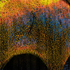
Gingiva is the soft tissue that surrounds and protects the teeth. Healthy gingiva provides an effective barrier to periodontal insults to deeper tissue, thus is an important indicator to a patient's periodontal health. Current methods in assessing gingival tissue health, including visual observation and physical examination with probing on the gingiva, are qualitative and subjective. They may become cumbersome when more complex cases are involved, such as variations in gingival biotypes where feature and thickness of the gingiva are considered. A noninvasive imaging technique providing depth-resolved structural and vascular information is necessary for an improved assessment of gingival tissue and more accurate diagnosis of periodontal status. We propose a three-dimensional (3D) imaging technique, optical coherence tomography (OCT), to perform in situ imaging on human gingiva. Ten volunteers (five male, five female, age 25-35) were recruited; and the labial gingival tissues of upper incisors were scanned using the combined use of state-of-the-art swept-source OCT and OCT angiography (OCTA). Information was collected describing the 3D tissue microstructure and capillary vasculature of the gingiva within a penetration depth of up to 2 mm. Results indicate significant structural and vascular differences between the two extreme gingival biotypes (ie, thick and thin gingiva), and demonstrate special features of vascular arrangement and characteristics in gingival inflammation. Within the limit of this study, the OCT/OCTA technique is feasible in quantifying different attributes of gingival biotypes and the severity of gingival inflammation.
image

The full text of this article hosted at iucr.org is unavailable due to technical difficulties.