Whole-Cell Lysosome SMLM Imaging as Indicators for Functional Diagnostics with a Low-Phototoxic Spontaneously Blinking Probe
Graphical Abstract
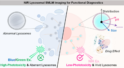
An NIR spontaneously blinking fluorophore, Aze-HMSiR, optimized for long-term SMLM imaging of lysosomes is developed. It exhibits superior photostability, pH responsiveness, and minimal phototoxicity. Aze-HMSiR can reveal key lysosomal characteristics, including distribution, size, and lumen pH, in response to various anticancer agents. Such whole-cell lysosome SMLM imaging serves as a valuable indicator for lysosomal functional diagnostics.
Abstract
Lysosomal morphology and pH dynamics are key indicators of lysosomal function, making long-term single-molecule localization microscopy (SMLM) imaging a promising tool for functional diagnostics. However, phototoxicity often compromises imaging reliability. Here, we develop Aze-HMSiR, a spontaneously blinking silicon rhodamine probe with near-infrared excitation, enabling low-phototoxicity, long-term (50 min) SMLM imaging of lysosomal morphology and pH dynamics. This probe provides super-resolution insights into lysosomal distribution, size, and lumen pH, essential for understanding their physiological and pathological roles. Using Aze-HMSiR, we investigate lysosomal function under acidosis, starvation, and anticancer drug treatments. Among seven tested drugs, periplocoside significantly reduced lysosomal size, while paclitaxel increased pH and altered lysosomal distribution. These findings highlight Aze-HMSiR as a powerful tool for lysosomal functional diagnostics and drug screening, offering a robust platform for studying lysosomal dynamics in response to physiological and pharmacological perturbations, particularly in cancer therapy.
Conflict of Interests
The authors declare no conflict of interest.
Open Research
Data Availability Statement
The data that support the findings of this study are available from the corresponding author upon reasonable request.


