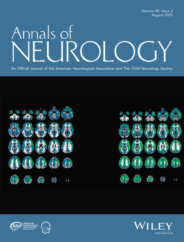
Tertiary microvascular territories define lacunar infarcts in the basal ganglia
Joel A. Feekes BA
The University of Iowa, Neuroscience PhD Program, University of Iowa Hospitals and Clinics, Iowa City, IA
Search for more papers by this authorShih-Wei Hsu MD
Department of Radiology, Interventional Neuroradiology Service, University of Iowa Hospitals and Clinics, Iowa City, IA
Section of Neuroradiology, Department of Diagnostic Radiology, Chang Gung Memorial Hospital, Kaohsiung Medical Center, Taiwan, Republic of China
Search for more papers by this authorJohn C. Chaloupka MD, FAHA, FACA
Department of Radiology, Interventional Neuroradiology Service, University of Iowa Hospitals and Clinics, Iowa City, IA
Search for more papers by this authorCorresponding Author
Martin D. Cassell PhD
Department of Anatomy and Cell Biology, University of Iowa, College of Medicine, Iowa City, IA
University of Iowa, Department of Anatomy and Cell Biology, 51 Newton Road, 1-500 Bowen Science Building, Iowa City, IA 52242-1109Search for more papers by this authorJoel A. Feekes BA
The University of Iowa, Neuroscience PhD Program, University of Iowa Hospitals and Clinics, Iowa City, IA
Search for more papers by this authorShih-Wei Hsu MD
Department of Radiology, Interventional Neuroradiology Service, University of Iowa Hospitals and Clinics, Iowa City, IA
Section of Neuroradiology, Department of Diagnostic Radiology, Chang Gung Memorial Hospital, Kaohsiung Medical Center, Taiwan, Republic of China
Search for more papers by this authorJohn C. Chaloupka MD, FAHA, FACA
Department of Radiology, Interventional Neuroradiology Service, University of Iowa Hospitals and Clinics, Iowa City, IA
Search for more papers by this authorCorresponding Author
Martin D. Cassell PhD
Department of Anatomy and Cell Biology, University of Iowa, College of Medicine, Iowa City, IA
University of Iowa, Department of Anatomy and Cell Biology, 51 Newton Road, 1-500 Bowen Science Building, Iowa City, IA 52242-1109Search for more papers by this authorAbstract
Lacunar infarcts are commonly found in the basal ganglia, though little is known about the organization of small-scale microvascular territories that presumably subtend lacunae. We investigated microvascular territories of the lenticulostriate arteries, the recurrent artery of Heubner, the anterior choroidal artery, and striate branches of the anterior cerebral and anterior communicating arteries in perfusion-fixed human brains by simultaneous injection of fluorescent dyes and a radio-opaque substance in 5% gelatin. Territories were defined by ultraviolet illumination of dye and high-resolution mammography of radio-opaque substance. Brains were sectioned coplanar with the Talairach proportional grid system and vascular data were plotted, allowing for application to any human brain. The data suggest first that the lenticulostriate artery, recurrent artery of Heubner, and anterior choroidal artery supply distinct territories of the basal ganglia with minimal overlap and sparse anastomoses between major penetrating vessels. Individual territories are spatially consistent across brains and match the extent of major/minor infarcts. Second, branching patterns of parental, second-, and third-order vessels leading to circumscribed terminal vascular beds could account structurally for “lacunar” infarcts. Ann Neurol 2005
References
- 1 Alexander L. The vascular supply of the striopallidum. Res Publ Assn Res Nerv Mental Disord 1942; 21: 77–132.
- 2 Gillilan L. The arterial and venous blood supplies to the forebrain (including the internal capsule) of primates. Neurology 1968; 18: 653–670.
- 3 Kaplan H. The lateral perforating branches of the anterior and middle cerebral arteries. J Neurosurg 1965; 23: 305–310.
- 4 Leeds N. The striate (lenticulostriate) arteries and the artery of Heubner. In: T Newton, D Potts, eds. Radiology of the skull and brain. Vol 2. St. Louis, MO: Mosby, 1974: 1527–1539.
- 5 Lin J, Kricheff I. The anterior cerebral artery complex. Section I. Normal anterior cerebral artery complex. In: T Newton, D Potts, eds. Radiology of the skull and brain. Vol 2. St. Louis, MO: Mosby, 1974: 1391–1396.
- 6 Perlmutter D, Rhoton A. Microsurgical anatomy of the distal anterior cerebral artery. J Neurosurg 1978; 49: 204–228.
- 7 Shellshear J. The basal arteries of the forebrain and their functional significance. J Anat 1920; 55: 27–35.
- 8 Ahmed D, Ahmed R. The recurrent branch of the anterior cerebral artery. Anatomical Records 1967; 157: 699–700.
- 9 Buchanan A. Functional neuro-anatomy. 4th ed. Philadelphia: Lea & Febiger Publishing, 1961.
- 10 Critchley M. The anterior cerebral artery and its syndromes. Brain 1953; 30: 120–165.
- 11 Gomes F, Dujovny M, Umansky F, et al. Microsurgical anatomy of the recurrent artery of Heubner. J Neurosurg 1984; 60: 130–139.
- 12 Kakou M, Destrieux C, Velut S. Microanatomy of the pericallosal arterial complex. J Neurosurg 2000; 93: 667–675.
- 13 Ostrowski AZ, Webster JE, Gurdjian ES. The proximal anterior cerebral artery: an anatomic study. Arch Neurol 1960; 3: 661–664.
- 14 Perlmutter D, Rhoton A. Microsurgical anatomy of the anterior cerebral-anterior communicating-recurrent artery complex. J Neurosurg 1976; 45: 259–272.
- 15 Rhoton A, Saeki N, Perlmutter D, Zeal A. Microsurgical anatomy of common aneurysm sites. Clin Neurosurg 1979; 26: 248–306.
- 16 Rosner S, Rhoton A, Ono M, Barry M. Microsurgical anatomy of the anterior perforating arteries. J Neurosurg 1984; 61: 468–485.
- 17
Rubinstein H.
Relation of circulus arteriosus to hypothalamus and internal capsule.
Arch Neurol Psychiatry
1944;
52:
526–530.
10.1001/archneurpsyc.1944.02290360098009 Google Scholar
- 18 Takahashi S, Goto K, Fukasawa H, et al. Computed tomography of cerebral infarction along the distribution of the basal perforating arteries. Part 1: striate arterial group. Radiology 1985; 155: 107–118.
- 19 Takahashi S, Goto K, Fukasawa H, et al. Computed tomography of cerebral infarction along the distribution of the basal perforating arteries. Part II: thalamic arterial group. Radiology 1985; 155: 119–130.
- 20 Westberg G. The recurrent artery of Heubner and the arteries of the central ganglia. Acta Radiol Diagn (Stockh) 1963; 11: 949–954.
- 21 Abbie A. The morphology of the forebrain arteries, with especial reference to the evolution of the basal ganglia. J Anat 1934; 68: 433–470.
- 22 Ghika J, Bogousslavsky J, Regli F. Infarcts in the territory of lenticulostriate branches from the middle cerebral artery: etiological factors and clinical features in 65 cases. Archives Suisses de Neurologie et de Psychiatrie 1991; 142: 5–18.
- 23 Marinkovic R, Markovic L. The arterial supply of the human amygdaloid body. Medicinski Pregled 1991; 44: 229–230.
- 24 Marinkovic S, Gibo H, Milisavljevic M, Cetkovic M. Anatomic and clinical correlations of the lenticulostriate arteries. Clin Anat 2001; 14: 190–195.
- 25 Marinkovic S, Milisavljevic M, Kovacevic M, Stevic Z. Perforating branches of the middle cerebral artery: microanatomy and clinical significance of their intracerebral segments. Stroke 1985; 16: 1022–1029.
- 26 Peele T. The neuroanatomical basis for clinical neurology. New York: McGraw-Hill Book Company, 1954.
- 27 Donzelli R, Marinkovic S, Brigante L, et al. Territories of the perforating (lenticulostriate) branches of the middle cerebral artery. Surg Radiol Anat 1998; 20: 393–398.
- 28 Grand W. Microsurgical anatomy of the proximal middle cerebral artery and the internal carotid artery bifurcation. Neurosurgery 1980; 7: 215–218.
- 29 Ture U, Yasargil M, Al-Mefty O, et al. Arteries of the insula. J Neurosurg 2000; 92: 676–687.
- 30 Abbie A. The clinical significance of the anterior choroidal artery. Brain 1933; 56: 233–246.
- 31 Carpenter M, Noback C, Moss M. The anterior choroidal artery: its origins, course, distribution and variations. Arch Neurol Psychiatry 1954; 71: 714–722.
- 32 Goldberg H. The anterior choroidal artery. In: T Newton, D Potts, eds. Radiology of the skull and brain. Vol 2. St. Louis, MO: Mosby, 1974: 1628–1658.
- 33 Helgason C, Caplan L, Goodwin J, Hedges T. Anterior choroidal artery-territory infarction: report of cases and review. Arch Neurol 1986; 43: 681–686.
- 34 Herman L, Fernando O, Gurdjian E. The anterior choroidal artery: an anatomical study of its area of distribution. Anatomical Records 1966; 154: 95–102.
- 35 Rhoton A, Fujii K, Fradd B. Microsurgical anatomy of the anterior choroidal artery. Surg Neurol 1979; 12: 171–187.
- 36 Theron J, Newton T. Anterior choroidal artery. I. Anatomic and radiographic study. J Neuroradiol 1976; 3: 5–30.
- 37 Westberg G. Arteries of the basal ganglia. Acta Radiol Diagn (Stockh) 1966; 5: 581–596.
- 38 Zeal AA, Rhoton AL Jr. Microsurgical anatomy of the posterior cerebral artery. J Neurosurg 1978; 48: 534–559.
- 39 Fisher C. Concerning strokes. Can Med Assoc J 1953; 69: 257–268.
- 40 Fisher C. Lacunes: small, deep cerebral infarcts. Neurology 1965; 15: 774–784.
- 41 Fisher CM. Lacunar strokes and infarcts: a review. Neurology 1982; 32: 871–876.
- 42 Fisher CM. Capsular infarcts: the underlying vascular lesions. Arch Neurol 1979; 36: 65–73.
- 43 Fisher CM. Lacunar infarcts—a review. Cerebrovasc Dis 1991; 1: 311–320.
- 44 Wardlaw JM, Dennis MS, Warlow CP, Sandercock PA. Imaging appearance of the symptomatic perforating artery in patients with lacunar infarction: occlusion or other vascular pathology? Ann Neurol 2001; 50: 208–215.
- 45 Fisher C. The arterial lesions underlying lacunes. Acta Neuropathologica (Berlin) 1969; 12: 1–15.
- 46 Lammie GA, Brannan F, Wardlaw JM. Incomplete lacunar infarction (type Ib lacunes). Acta Neuropathol (Berl) 1998; 96: 163–171.
- 47 Futrell N. Lacunar infarction: embolism is the key. Stroke 2004; 35: 1778–1779.
- 48 Norrving B. Lacunar infarction: embolism is the key: against. Stroke 2004; 35: 1779–1780.
- 49 Davis SM, Donnan GA. Why lacunar syndromes are different and important. Stroke 2004; 35: 1780–1781.
- 50 Ogata J. The arterial lesions underlying cerebral infarction. Neuropathology 1999; 19: 112–118.
- 51 Russmann H, Vingerhoets F, Ghika J, et al. Acute infarction limited to the lenticular nucleus: clinical, etiologic, and topographic features. Arch Neurol 2003; 60: 351–355.
- 52 Bhatia K, Marsden C. The behavioral and motor consequences of focal lesions of the basal ganglia in man. Brain 1994; 117: 859–876.
- 53 Thron A. Vascular anatomy of the spinal cord: neuroradiological investigations and clinical syndromes. New York: Springer-Verlag, 1945.
- 54 Talairach J, Tournoux P. Co-planar stereotaxic atlas of the human brain. New York: Thieme Medical, 1988.
- 55 Mai J, Assheuer J, Paxinos G. Atlas of the human brain. San Diego, CA: Academic Press, 1997.
- 56
Duvernoy H.
The human hippocampus: an atlas of applied anatomy.
Munich, Germany:
JF Bergmann Verlag,
1988.
10.1007/978-3-642-54195-7 Google Scholar
- 57 Marinkovic S, Kovacevic M, Marinkovic J. Perforating branches of the middle cerebral artery: microsurgical anatomy of their extracerebral segments. J Neurosurg 1985; 63: 266–271.
- 58 Hsieh F, Cheng T. Distribution of cerebral infarcts on computed tomography. Kaohsiung J Med Sci 1990; 6: 523–528.
- 59 Marinkovic S, Milisavljevic M, Marinkovic Z. Branches of the anterior communicating artery: microsurgical anatomy. Acta Neurochirurgica 1990; 106: 78–85.
- 60 Challa VR, Bell MA, Moody DM. A combined hematoxylin-eosin, alkaline phosphatase and high-resolution microradiographic study of lacunes. Clin Neuropathol 1990; 9: 196–204.
- 61 Lewis OJ. The form and development of the blood vessels of the mammalian cerebral cortex. J Anat 1957; 91: 40–46.
- 62 Brown L, Breuer O, Smith D, Lawhorn C. The striosomal labyrinth of the striatum in mice and its relationship to the organization and density of blood vessels: more evidence for a lattice-like functional organization in the striatum. Washington, DC: Society for Neuroscience, 2004.
- 63 Brown LL, Feldman SM, Smith DM, et al. Differential metabolic activity in the striosome and matrix compartments of the rat striatum during natural behaviors. J Neurosci 2002; 22: 305–314.


