Digital Cytology Combined With Artificial Intelligence Compared to Conventional Microscopy for Anal Cytology: A Preliminary Study
Funding: The authors received no specific funding for this work.
ABSTRACT
Introduction
Recent studies have shown that digital cytology (DC) coupled with artificial intelligence (AI) algorithms is a valid approach to the diagnosis of cervico-vaginal lesions using liquid-based cytology (LBC). We evaluated the use of these methods for anal LBC specimens.
Methods
A series of 124 anal LBC slides previously diagnosed by conventional microscopy (CC) were reviewed with a DC/AI system that generated a gallery of images. Diagnoses based on the selected images, according to the 2014 Bethesda System for Reporting Cervical Cytology, were compared to CC.
Results
Overall, CC and DC/AI approaches detected a similar number of abnormal (ASC-US+) cases (63 and 62 cases, respectively). We observed an exact concordance between CC and DC in 70 (57.9%) cases, corresponding to a moderate agreement between the two approaches (κ = 0.41, p < 0.001). A moderate agreement (κ = 0.48, p < 0.001) was also found when positive cases were stratified into ‘low-grade’ (ASC-US, LSIL) and ‘high-grade’ lesions (ASC-H, HSIL). The DC/AI system detected more cases of higher severity (ASC-H, HSIL: 9 and 2 cases, respectively) than CC (3 cases classified as HSIL).
Conclusions
The number of ASC-US+ cases detected by both systems was similar. The DC/AI system detected more cases of higher severity compared to the CC.
Graphical Abstract
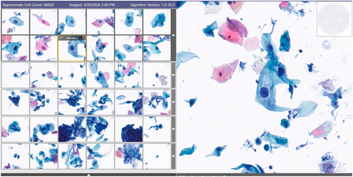
Cytology is an accepted method of screening for anal cancer. We compared the diagnostic performance between conventional microscopy and digital cytology in a series of anal specimens. The number of ASC-US+ cases detected by both systems was similar. However, the digital cytology system detected more high-grade lesions than conventional microscopy.
This study compared the diagnostic performance between conventional microscopy (CC) and digital cytology (DC) coupled with artificial intelligence (AI) in a series of anal cytology specimens. Although the number of ASC-US+ cases detected by both systems was similar, the DC/AI system detected more high-grade lesions than CC.
Conflicts of Interest
The authors declare no conflicts of interest.
Open Research
Data Availability Statement
The data that support the findings of this study are available from the corresponding author upon reasonable request.


