Fine-Needle Aspiration Biopsy of Adrenal Gland Lesions: The Roles of Image Guidance, Rapid On-Site Evaluation and Additional Tissue Sampling
Funding: The authors received no specific funding for this work.
This work was presented in part at the 67th Annual Scientific Meeting of American Society of Cytopathology, Salt Lake City, Utah, 14–17 November 2019.
ABSTRACT
Objective
An accurate fine-needle aspiration (FNA) diagnosis of adrenal lesions may be challenging. This study was to investigate roles of imaging guidance, rapid on-site evaluation (ROSE) and additional tissue sampling in FNA diagnosis of adrenal lesions.
Methods
Adrenal FNA cases were retrieved from pathology archive. Patients' demographics, lesion size and location, imaging guidance methods, cytologic diagnoses and histopathologic diagnoses were reviewed and analysed.
Results
The study cohort included 72 cases of left (86%) and right (14%) adrenal lesions. Endoscopic ultrasound (EUS) and computed tomography (CT) were used in 47 (65%) and 25 (35%) cases, respectively. Left adrenal lesions were sampled mostly by EUS-FNA (73%), whereas right adrenal lesions by CT-guided FNA (80%). There were no differences between the EUS-FNA and CT-FNA groups in terms of non-diagnostic rate and cytologic diagnostic categories. The non-diagnostic rate and cytologic diagnostic categories were the same between ROSE and non-ROSE groups. In a subset of 18 cases with concurrent core tissue biopsy, a definite diagnosis was rendered in all biopsy cases including three cases with a non-diagnostic or indeterminate cytology diagnosis.
Conclusion
Our study demonstrates that FNA has great efficacy for evaluation of adrenal lesions, either via EUS or CT guidance. Incorporation of ROSE evaluation into FNA procedure does not directly affect the performance of FNA biopsy but may help direct additional tissue sampling to salvage the cases with a non-diagnostic or indeterminate cytology diagnosis, increasing diagnostic yield.
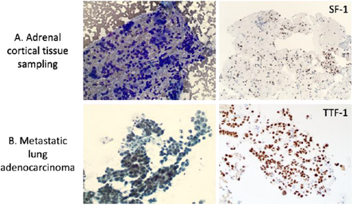
Graphical Abstract
This study demonstrates that fine-needle aspiration (FNA) has great efficacy for evaluation of adrenal lesions, either under endoscopic ultrasound or CT guidance. Incorporation of rapid on-site evaluation into FNA procedure does not directly affect the performance of FNA biopsy but may help direct additional tissue sampling to increase diagnostic yield.
Conflicts of Interest
The authors declare no conflicts of interest.
Open Research
Data Availability Statement
The data that support the findings of this study are available from the corresponding author upon request.


