Squash smear and fine needle aspiration features of conventional chordoma
Abstract
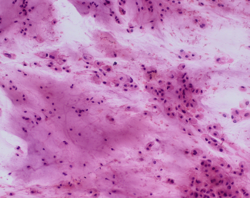
Chordoma is a rare primary central nervous system tumour of notochordal origin. Proper intraoperative or preoperative diagnosis of this entity is crucial for appropriate surgical management. The most common histopathological subtype is conventional chordoma. Cytological characteristics of this subtype are quite distinctive and the diagnosis can be easily made by cytology. There are two particularly important features that are observed in both squash smear and fine needle aspiration specimens: an abundant myxochondroid stroma and cells with large vacuoles, including physaliferous cells. The main differential diagnosis is conventional chondrosarcoma, but in problematic cases immunohistochemical studies are useful to establish the correct diagnosis.
Graphical Abstract
A proper intraoperative or preoperative diagnosis of conventional chordoma is crucial for appropriate surgical management because incomplete excision reduces the likelihood of progression-free survival and overall survival. The article presents the characteristic cytological features (including an abundant myxochondroid stroma and cells with large vacuoles) and specific immunohistochemical staining results (nuclear brachyury positivity and co-expression of epithelial markers and S100) that make cytology an effective tool for the diagnosis of this entity.
CONFLICT OF INTEREST STATEMENT
The authors declare no conflict of interest.
Open Research
DATA AVAILABILITY STATEMENT
Data sharing not applicable.


