Morphology quiz: Bronchial washing cytology from flexible bronchoscopy
Abstract
The aim of this observation is to make cytologists aware of the identification of melanoma cells in bronchial washings from an endobronchial metastasis of malignant melanoma. CT scan and flexible bronchoscopy images are provided and differential diagnosis and additional analyses (molecular biology) are mentioned and discussed.
1 CASE HISTORY
- 75-year-old man
- Former smoker
- History of right scapular malignant melanoma (Clark level IV; Breslow thickness 4 mm) treated by local surgery 7 years ago
- Current clinical presentation: Local recurrence at the right scapular scar
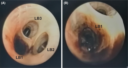
- Paraclinical findings: Imagery showed a right dorsal subcutaneous tumour mass of 58 mm associated with right axillary adenopathies and proximal endobronchial obstruction of the apical segment of the left upper lobe (LB1) (Figures 1, 2). Flexible bronchoscopy was performed highlighting a dark pigmented endobronchial tumour in LB1 (Figure 2B) and bronchial washing was collected for cytological analysis (Figure 3).


2 MORPHOLOGY QUIZ
- What is the most frequent origin of endobronchial tumour?
- Lung
- Breast
- Cutaneous melanoma
- Kidney
- Which cells are usually observed in a representative bronchial washing?
- Macrophages
- Bronchial cells
- Superficial Malpighian cells
- Mesothelial cells
- Based on the images provided in Figure 3, what is the likely cytological diagnosis of the dyskaryotic and dark pigmented cells?
- Adenocarcinoma
- Squamous cell carcinoma
- Malignant melanoma
- Lymphoma
- What is the most pertinent marker to search by molecular biology for malignant melanoma treatment optimisation?
- ROS1 rearrangement
- BRAF p.V600E mutation
- RAS p.G12D mutation
- EGFR exon 19 deletion
AUTHOR CONTRIBUTIONS
ANSWERS TO THE MULTIPLE-CHOICE QUESTIONS
- a. Comments: Endobronchial metastasis occurs in less than 5% of extrapulmonary malignancies.1, 2
- a and b. Comments: Superficial Malpighian cells from oropharynx are ausual contaminant of cytological samples obtained from a bronchoscopy procedure. A bronchial washing containing a majority of superficial Malpighian cells is considered not representative.3
- c. Comments: (1) Diagnosis of metastatic endobronchial melanoma was confirmed on lung biopsy, showing positivity of malignant cells with SOX10 marker. (2) “Black bronchoscopy” corresponding to dark pigmentation of the endobronchial tree can have several aetiologies: neoplasms but also congenital causes, environmental causes, iatrogenic causes.4
- b. Comments: BRAF V600E mutation was identified from lung biopsy.
ACKNOWLEDGEMENTS
None. Open access funding enabled and organized by ProjektDEAL.
CONFLICT OF INTEREST
The authors have no conflicts of interest to declare.
Open Research
DATA AVAILABILITY STATEMENT
Data sharing is not applicable to this article as no new data were created or analyzed in this study.


