Volume 13, Issue 2 e202070005
COVER PICTURE
Free Access

Inside Cover: Optical coherence tomography as a noninvasive 3D real time imaging tool for the rapid evaluation of phenotypic variations in insect embryonic development (J. Biophotonics 2/2020)

Ya Su

Ya Su

Photonics Information Innovation Center, Hebei Provincial Center for Optical Sensing Innovations, College of Physics Science & Technology, Hebei University, Baoding, China

Search for more papers by this author
Liya Wei

Liya Wei

College of Life Sciences, Hebei University, Baoding, China

Search for more papers by this author
Hao Tan

Hao Tan

Photonics Information Innovation Center, Hebei Provincial Center for Optical Sensing Innovations, College of Physics Science & Technology, Hebei University, Baoding, China

Search for more papers by this author
Jing Li

Jing Li

College of Life Sciences, Hebei University, Baoding, China

Search for more papers by this author
Wenping Li

Wenping Li

Photonics Information Innovation Center, Hebei Provincial Center for Optical Sensing Innovations, College of Physics Science & Technology, Hebei University, Baoding, China

Search for more papers by this author
Lei Fu

Lei Fu

Photonics Information Innovation Center, Hebei Provincial Center for Optical Sensing Innovations, College of Physics Science & Technology, Hebei University, Baoding, China

Search for more papers by this author
Tongxin Wang

Tongxin Wang

College of Life Sciences, Hebei University, Baoding, China

Search for more papers by this author
Le Kang

Corresponding Author

Le Kang

College of Life Sciences, Hebei University, Baoding, China

Correspondence

X. Steve Yao, Photonics Information Innovation Center, Hebei Provincial Center for Optical Sensing Innovations, College of Physics Science & Technology, Hebei University, Baoding 071002, China.

Email: [email protected]

Le Kang, College of Life Sciences, Hebei University, Baoding 071002, China.

Email: [email protected]

Search for more papers by this author
X. Steve Yao

Corresponding Author

X. Steve Yao

Photonics Information Innovation Center, Hebei Provincial Center for Optical Sensing Innovations, College of Physics Science & Technology, Hebei University, Baoding, China

Correspondence

X. Steve Yao, Photonics Information Innovation Center, Hebei Provincial Center for Optical Sensing Innovations, College of Physics Science & Technology, Hebei University, Baoding 071002, China.

Email: [email protected]

Le Kang, College of Life Sciences, Hebei University, Baoding 071002, China.

Email: [email protected]

Search for more papers by this author
First published: 03 February 2020
Citations: 1

Abstract

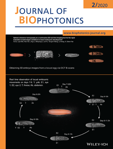
Top: Illustration of how to obtain 3D embryo images from a locust egg using OCT B-scans. The internal structures of the embryo can be clearly identified as it develops.

Bottom: The real time observation of katatrepsis and twist of a lowland locust embryo on days 7-8, as the embryo develops. The embryonic movements can be readily identified by tracking the positions of the embryo's eyes (E1 and E2).

Further details can be found in the article by Ya Su, Liya Wei, Hao Tan, et al. (e201960047).

The full text of this article hosted at iucr.org is unavailable due to technical difficulties.