Visibility of microvessels in Optical Coherence Tomography angiography depends on angular orientation
Funding information: National Institutes of Health, Grant/Award Numbers: R01EY028287, R01EY030361, R01EY031469, R01NS094681, R01EB029747, R21NS105043
Abstract
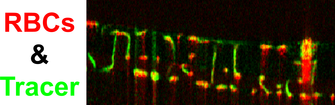
Optical Coherence Tomography angiography (OCTA) is a widespread tool for depth-resolved imaging of chorioretinal vasculature with single microvessel resolution. To improve the clinical interpretation of OCTA, the conditions affecting visualization of microvessels must be defined. Here we inject a scattering plasma tracer (Intralipid) during OCTA imaging of the anesthetized rat eye. In the retina, we find that interlaminar (vertical) vessels that connect laminae have one-fourth to one-third the OCTA red blood cell to tracer (RBC-to-tracer) signal ratio of intralaminar (horizontal) vessels. This finding suggests that the OCTA signal from microvessels depends on angular orientation, making vertically-oriented vessels more difficult to visualize using intrinsic contrast alone. Clinicians should be aware of this potential artifact when interpreting OCTA.