Volume 13, Issue 1 e201960062
FULL ARTICLE

Automated label-free detection of injured neuron with deep learning by two-photon microscopy

Shu Wang

Shu Wang

College of Mechanical Engineering and Automation, Fuzhou University, Fuzhou, China

Search for more papers by this author
Bingbing Lin

Bingbing Lin

College of Rehabilitation Medicine, Fujian University of Traditional Chinese Medicine, Fuzhou, China

Search for more papers by this author
Guimin Lin

Guimin Lin

College of Physics & Electronic Information Engineering, Minjiang University, Fuzhou, China

Search for more papers by this author
Ruolan Lin

Ruolan Lin

Department of Radiology, Fujian Medical University Union Hospital, Fuzhou, China

Search for more papers by this author
Feng Huang

Feng Huang

College of Mechanical Engineering and Automation, Fuzhou University, Fuzhou, China

Search for more papers by this author
Weilin Liu

Weilin Liu

College of Rehabilitation Medicine, Fujian University of Traditional Chinese Medicine, Fuzhou, China

Search for more papers by this author
Xingfu Wang

Xingfu Wang

Department of Pathology, The First Affiliated Hospital of Fujian Medical University, Fuzhou, China

Search for more papers by this author
Xueyong Liu

Xueyong Liu

Department of Pathology, The First Affiliated Hospital of Fujian Medical University, Fuzhou, China

Search for more papers by this author
Yu Zhang

Yu Zhang

Department of Pathology, The First Affiliated Hospital of Fujian Medical University, Fuzhou, China

Search for more papers by this author
Feng Wang

Feng Wang

Department of Neurosurgery, The First Affiliated Hospital of Fujian Medical University, Fuzhou, China

Search for more papers by this author
Yuanxiang Lin

Yuanxiang Lin

Department of Neurosurgery, The First Affiliated Hospital of Fujian Medical University, Fuzhou, China

Search for more papers by this author
Lidian Chen

Corresponding Author

Lidian Chen

College of Rehabilitation Medicine, Fujian University of Traditional Chinese Medicine, Fuzhou, China

Correspondence

Lidian Chen, College of Rehabilitation Medicine, Fujian University of Traditional Chinese Medicine, Fuzhou 350122, China.

Email: [email protected]

Jianxin Chen, Key Laboratory of OptoElectronic Science and Technology for Medicine of Ministry of Education, Fujian Provincial Key Laboratory of Photonics Technology, Fujian Normal University, Fuzhou 350007, China.

Email: [email protected]

Search for more papers by this author
Jianxin Chen

Corresponding Author

Jianxin Chen

Key Laboratory of OptoElectronic Science and Technology for Medicine of Ministry of Education, Fujian Provincial Key Laboratory of Photonics Technology, Fujian Normal University, Fuzhou, China

Correspondence

Lidian Chen, College of Rehabilitation Medicine, Fujian University of Traditional Chinese Medicine, Fuzhou 350122, China.

Email: [email protected]

Jianxin Chen, Key Laboratory of OptoElectronic Science and Technology for Medicine of Ministry of Education, Fujian Provincial Key Laboratory of Photonics Technology, Fujian Normal University, Fuzhou 350007, China.

Email: [email protected]

Search for more papers by this author
First published: 11 October 2019
Citations: 13
Shu Wang, Bingbing Lin and Guimin Lin contributed equally to this study.

Funding information: the Program for Changjiang Scholars and Innovative Research Team in University, Grant/Award Number: IRT_15R10; National High Technology Research and Development Program of China, Grant/Award Number: 2015AA020508; National Key Basic Research Program of China, Grant/Award Number: 2015CB352006; National Natural Science Foundation of China, Grant/Award Numbers: 61972187, 81671730; Natural Science Foundation of Fujian Province of China, Grant/Award Number: 2019J01761; Program for Undergraduate Education and Teaching Reform in University of Fujian Province of China, Grant/Award Number: FBJG20190230; the Joint Funds of Fujian Provincial Health and Education Research, Grant/Award Number: WKJ2016-2-28; the Special Funds of the Central Government Guiding Local Science and Technology Development, Grant/Award Number: 2017L3009

Abstract

Stroke is a significant cause of morbidity and long-term disability globally. Detection of injured neuron is a prerequisite for defining the degree of focal ischemic brain injury, which can be used to guide further therapy. Here, we demonstrate the capability of two-photon microscopy (TPM) to label-freely identify injured neurons on unstained thin section and fresh tissue of rat cerebral ischemia-reperfusion model, revealing definite diagnostic features compared with conventional staining images. Moreover, a deep learning model based on convolutional neural network is developed to automatically detect the location of injured neurons on TPM images. We then apply deep learning-assisted TPM to evaluate the ischemic regions based on tissue edema, two-photon excited fluorescence signal intensity, as well as neuronal injury, presenting a novel manner for identifying the infarct core, peri-infarct area, and remote area. These results propose an automated and label-free method that could provide supplementary information to augment the diagnostic accuracy, as well as hold the potential to be used as an intravital diagnostic tool for evaluating the effectiveness of drug interventions and predicting potential therapeutics.image

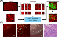
CONFLICT OF INTEREST

The authors declare no financial or commercial conflict of interest.

The full text of this article hosted at iucr.org is unavailable due to technical difficulties.