Pathological crystal imaging with single-shot computational polarized light microscopy
Bijie Bai
Electrical and Computer Engineering Department, University of California, Los Angeles, Los Angeles, California
Bioengineering Department, University of California, Los Angeles, Los Angeles, California
California NanoSystems Institute, University of California, Los Angeles, Los Angeles, California
Search for more papers by this authorHongda Wang
Electrical and Computer Engineering Department, University of California, Los Angeles, Los Angeles, California
Bioengineering Department, University of California, Los Angeles, Los Angeles, California
California NanoSystems Institute, University of California, Los Angeles, Los Angeles, California
Search for more papers by this authorTairan Liu
Electrical and Computer Engineering Department, University of California, Los Angeles, Los Angeles, California
Bioengineering Department, University of California, Los Angeles, Los Angeles, California
California NanoSystems Institute, University of California, Los Angeles, Los Angeles, California
Search for more papers by this authorYair Rivenson
Electrical and Computer Engineering Department, University of California, Los Angeles, Los Angeles, California
Bioengineering Department, University of California, Los Angeles, Los Angeles, California
California NanoSystems Institute, University of California, Los Angeles, Los Angeles, California
Search for more papers by this authorJohn FitzGerald
Division of Rheumatology, Department of Internal Medicine, David Geffen School of Medicine, University of California, Los Angeles, Los Angeles, California
Search for more papers by this authorCorresponding Author
Aydogan Ozcan
Electrical and Computer Engineering Department, University of California, Los Angeles, Los Angeles, California
Bioengineering Department, University of California, Los Angeles, Los Angeles, California
California NanoSystems Institute, University of California, Los Angeles, Los Angeles, California
Department of Surgery, David Geffen School of Medicine, University of California, Los Angeles, Los Angeles, California
Correspondence
Aydogan Ozcan, University of California, Los Angeles, CA 90095.
Email: [email protected]
Search for more papers by this authorBijie Bai
Electrical and Computer Engineering Department, University of California, Los Angeles, Los Angeles, California
Bioengineering Department, University of California, Los Angeles, Los Angeles, California
California NanoSystems Institute, University of California, Los Angeles, Los Angeles, California
Search for more papers by this authorHongda Wang
Electrical and Computer Engineering Department, University of California, Los Angeles, Los Angeles, California
Bioengineering Department, University of California, Los Angeles, Los Angeles, California
California NanoSystems Institute, University of California, Los Angeles, Los Angeles, California
Search for more papers by this authorTairan Liu
Electrical and Computer Engineering Department, University of California, Los Angeles, Los Angeles, California
Bioengineering Department, University of California, Los Angeles, Los Angeles, California
California NanoSystems Institute, University of California, Los Angeles, Los Angeles, California
Search for more papers by this authorYair Rivenson
Electrical and Computer Engineering Department, University of California, Los Angeles, Los Angeles, California
Bioengineering Department, University of California, Los Angeles, Los Angeles, California
California NanoSystems Institute, University of California, Los Angeles, Los Angeles, California
Search for more papers by this authorJohn FitzGerald
Division of Rheumatology, Department of Internal Medicine, David Geffen School of Medicine, University of California, Los Angeles, Los Angeles, California
Search for more papers by this authorCorresponding Author
Aydogan Ozcan
Electrical and Computer Engineering Department, University of California, Los Angeles, Los Angeles, California
Bioengineering Department, University of California, Los Angeles, Los Angeles, California
California NanoSystems Institute, University of California, Los Angeles, Los Angeles, California
Department of Surgery, David Geffen School of Medicine, University of California, Los Angeles, Los Angeles, California
Correspondence
Aydogan Ozcan, University of California, Los Angeles, CA 90095.
Email: [email protected]
Search for more papers by this authorFunding information: National Institutes of Health, Grant/Award Number: R21AR072946
Abstract
Pathological crystal identification is routinely practiced in rheumatology for diagnosing arthritis disease such as gout, and relies on polarized light microscopy as the gold standard method used by medical professionals. Here, we present a single-shot computational polarized light microscopy method that reconstructs the transmittance, retardance and slow-axis orientation of a birefringent sample using a single image captured with a pixelated-polarizer camera. This method is fast, simple-to-operate and compatible with all the existing standard microscopes without extensive or costly modifications. We demonstrated the success of our method by imaging three different types of crystals found in synovial fluid and reconstructed the birefringence information of these samples using a single image, without being affected by the orientation of individual crystals within the sample field-of-view. We believe this technique will provide improved sensitivity, specificity and speed, all at low cost, for clinical diagnosis of crystals found in synovial fluid and other bodily fluids.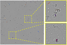
CONFLICT OF INTEREST
A.O., B.B., and H.W. have a pending patent application on the presented technique. The remaining authors declare that they have no conflict of interest.
REFERENCES
- 1P. Arun Gopinathan, G. Kokila, M. Jyothi, C. Ananjan, L. Pradeep, S. Humaira Nazir, Scientifica (Cairo) 2015, 2015, 802980.
- 2K. Király, M. M. Hyttinen, T. Lapveteläinen, M. Elo, I. Kiviranta, J. Dobai, L. Módis, H. J. Helminen, J. P. Arokoski, Histochem. J. 1997, 29, 317.
- 3A. Changoor, N. Tran-Khanh, S. Méthot, M. Garon, M. B. Hurtig, M. S. Shive, M. D. Buschmann, Osteoarthr. Cartil. 2011, 19, 126.
- 4J. Orberg, E. Baer, A. Hiltner, Connect. Tissue Res. 1983, 11, 285.
- 5L.-W. Jin, K. A. Claborn, M. Kurimoto, M. A. Geday, I. Maezawa, F. Sohraby, M. Estrada, W. Kaminksy, B. Kahr, Proc. Natl. Acad. Sci. USA 2003, 100, 15294.
- 6B. Vijaya, B. S. Dalal, Sunila, and G. V. Manjunath, Indian J. Pathol. Microbiol. 55, 170–174 (2012).
- 7G. G. Cornwell, W. L. Murdoch, R. A. Kyle, P. Westermark, P. Pitkänen, Am. J. Med 1983, 75, 618.
- 8G. Schapira, J.-C. Dreyfus, M. Joly, Nature 1952, 170, 494.
- 9D. C. Adams, L. P. Hariri, A. J. Miller, Y. Wang, J. L. Cho, M. Villiger, J. A. Holz, M. V. Szabari, D. L. Hamilos, R. Scott Harris, J. W. Griffith, B. E. Bouma, A. D. Luster, B. D. Medoff, M. J. Suter, Sci. Transl. Med. 2016, 8, 359ra131.
- 10M. C. Pierce, J. Strasswimmer, H. Park, B. Cense, J. F. de Boer, J. Biomed. Opt. 2004, 9, 287.
- 11S. L. Jacques, J. C. Ramella-Roman, K. Lee, J. Biomed. Opt. 2002, 7, 329.
- 12D. J. Mccarty, J. L. Hollander, Ann. Intern. Med. 1961, 54, 452.
- 13E. Pascual, E. Batlle-Gualda, A. Martínez, J. Rosas, P. Vela, Ann. Intern. Med. 1999, 131, 756.
- 14M. Wolman, J. Histochem. Cytochem. 1975, 23, 21.
- 15D. J. Mccarty, N. N. Kohn, J. S. Faires, Ann. Intern. Med. 1962, 56, 711.
- 16N. W. McGill, P. A. Dieppe, Ann. Rheum. Dis. 1991, 50, 558.
- 17R. A. Gatter, Arthritis Rheum. 1974, 17, 253.
- 18A. Rosenthal, L. Ryan, D. McCarty, Arthritis and Allied Conditions: A Textbook of Rheumatology, 14th ed., Lippincott Williams & Wilkins, Philadelphia, PA 2001, p. 2348.
- 19J. W. Park, D. J. Ko, J. J. Yoo, S. H. Chang, H. J. Cho, E. H. Kang, J. K. Park, Y. W. Song, Y. J. Lee, Korean J. Intern. Med. 2014, 29, 361.
- 20R. Oldenbourg, G. Mei, J. Microsc. 1995, 180, 140.
- 21R. D. Goldman, D. L. Spector, Live Cell Imaging: A Laboratory Manual, Cold Spring Harbor Laboratory Press, Cold Spring Harbor, NY 2005, p. 205.
- 22Y. Zhang, S. Y. C. Lee, Y. Zhang, D. Furst, J. Fitzgerald, A. Ozcan, Sci. Rep. 2016, 6, 28793.
- 23C. S. L. Chun, D. L. Fleming, E. J. Torok, Polarization-sensitive thermal imaging, in Automatic Object Recognition IV (International Society for Optics and Photonics), Orlando, FL 1994, Vol. 2234, pp. 275–286.
- 24G. P. Nordin, J. T. Meier, P. C. Deguzman, M. W. Jones, J. Opt. Soc. Am. A 1999, 16, 1168.
- 25N. Brock, B. T. Kimbrough, J. E. Millerd, A pixelated micropolarizer-based camera for instantaneous interferometric measurements, in Polarization Science and Remote Sensing V (International Society for Optics and Photonics), San Diego, CA 2011, Vol. 8160, p. 81600W.
- 26J. S. Tyo, D. L. Goldstein, D. B. Chenault, J. A. Shaw, Appl. Optics, AO 2006, 45, 5453.
- 27X. Tian, X. Tu, K. D. Croce, G. Yao, H. Cai, N. Brock, S. Pau, R. Liang, Biomed. Opt. Express, BOE 2019, 10, 1638.
- 28C. B. Kahn, J. L. Hollander, H. R. Schumacher, JAMA 1970, 211, 807.
- 29T. Yamazaki, Y. Maruyama, Y. Uesaka, M. Nakamura, Y. Matoba, T. Terada, K. Komori, Y. Ohba, S. Arakawa, Y. Hirasawa, Y. Kondo, J. Murayama, K. Akiyama, Y. Oike, S. Sato, T. Ezaki, Four-directional pixel-wise polarization CMOS image sensor using air-gap wire grid on 2.5-μm back-illuminated pixels, in 2016 IEEE International Electron Devices Meeting (IEDM) San Francisco, CA 2016, pp. 8.7.1-8.7.4.
- 30H. Hurwitz, R. C. Jones, J. Opt. Soc. Am. 1941, 31, 493.
- 31M. Shribak, R. Oldenbourg, Appl. Optics 2003, 42, 3009.
- 32S. O. Isikman, W. Bishara, S. Mavandadi, F. W. Yu, S. Feng, R. Lau, A. Ozcan, Proc. Natl. Acad. Sci. USA 2011, 108, 7296.
Citing Literature