Volume 11, Issue 8 e201700126
FULL ARTICLE

High-speed microscopy for in vivo monitoring of lymph dynamics

Mustafa Sarimollaoglu

Corresponding Author

Mustafa Sarimollaoglu

Arkansas Nanomedicine Center, University of Arkansas for Medical Sciences, Little Rock, Arkansas

Correspondence

Mustafa Sarimollaoglu and Vladimir Zharov, Arkansas Nanomedicine Center, University of Arkansas for Medical Sciences, Little Rock, AR 72205. Email: [email protected], [email protected]

Search for more papers by this author
Amanda J. Stolarz

Amanda J. Stolarz

Department of Pharmacology and Toxicology, University of Arkansas for Medical Sciences, Little Rock, Arkansas

Search for more papers by this author
Dmitry A. Nedosekin

Dmitry A. Nedosekin

Arkansas Nanomedicine Center, University of Arkansas for Medical Sciences, Little Rock, Arkansas

Search for more papers by this author
Brittney R. Garner

Brittney R. Garner

Department of Pharmacology and Toxicology, University of Arkansas for Medical Sciences, Little Rock, Arkansas

Search for more papers by this author
Terry W. Fletcher

Terry W. Fletcher

Department of Pharmacology and Toxicology, University of Arkansas for Medical Sciences, Little Rock, Arkansas

Search for more papers by this author
Ekaterina I. Galanzha

Ekaterina I. Galanzha

Arkansas Nanomedicine Center, University of Arkansas for Medical Sciences, Little Rock, Arkansas

Search for more papers by this author
Nancy J. Rusch

Nancy J. Rusch

Department of Pharmacology and Toxicology, University of Arkansas for Medical Sciences, Little Rock, Arkansas

Search for more papers by this author
Vladimir P. Zharov

Corresponding Author

Vladimir P. Zharov

Arkansas Nanomedicine Center, University of Arkansas for Medical Sciences, Little Rock, Arkansas

Correspondence

Mustafa Sarimollaoglu and Vladimir Zharov, Arkansas Nanomedicine Center, University of Arkansas for Medical Sciences, Little Rock, AR 72205. Email: [email protected], [email protected]

Search for more papers by this author
First published: 12 December 2017
Citations: 12
Funding information National Cancer Institute, Grant/Award number: NCI R21 1CA131164; National Institutes of Health, Grant/Award numbers: R01CA131164, R01EB017217; National Science Foundation, Grant/Award numbers: OIA 1457888, DBI 1556068; Translational Research Institute grant from UAMS; Rho Chi AFPE First Year Graduate Fellowship; PhRMA Predoctoral Fellowship; UAMS College of Medicine Pilot Grant

Abstract

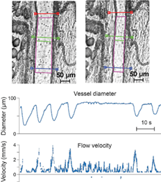
The lymphatic system contributes to body homeostasis by clearing fluid, lipids, plasma proteins and immune cells from the interstitial space. Many studies have been performed to understand lymphatic function under normal conditions and during disease. Nevertheless, a further improvement in quantification of lymphatic behavior is needed. Here, we present advanced bright-field microscopy for in vivo imaging of lymph vessels (LVs) and automated quantification of lymphatic function at a temporal resolution of 2 milliseconds. Full frame videos were compressed and recorded continuously at up to 540 frames per second. A new edge detection algorithm was used to monitor vessel diameter changes across multiple cross sections, while individual cells in the LVs were tracked to estimate flow velocity. The system performance initially was verified in vitro using 6- and 10-μm microspheres as cell phantoms on slides and in 90-μm diameter tubes at flow velocities up to 4 cm/second. Using an in vivo rat model, we explored the mechanisms of lymphedema after surgical lymphadenectomy of the mesentery. The system revealed reductions of mesenteric LV contraction and flow rate. Thus, the described imaging system may be applicable to the study of lymphatic behavior during therapeutic and surgical interventions, and potentially during lymphatic system diseases.

image

The full text of this article hosted at iucr.org is unavailable due to technical difficulties.