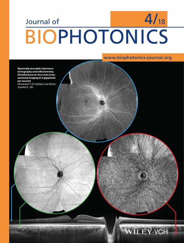
Pixel classification method in optical coherence tomography for tumor segmentation and its complementary usage with OCT microangiography
Corresponding Author
Alexander Moiseev
Nano-optics and Highly Sensitive Optical Measurement Department, Institute of Applied Physics Russian Academy of Sciences, Nizhny Novgorod, Russia
Laboratory of Optical Coherent Tomography, Nizhny Novgorod State Medical Academy, Nizhny Novgorod, Russia
Correspondence
Alexander Moiseev, Institute of Applied Physics Russian Academy of Sciences, Ulyanova Street 46, 603950 Nizhny Novgorod, Russia. Email: [email protected]
Search for more papers by this authorLudmila Snopova
Laboratory of Optical Coherent Tomography, Nizhny Novgorod State Medical Academy, Nizhny Novgorod, Russia
Search for more papers by this authorSergey Kuznetsov
Laboratory of Optical Coherent Tomography, Nizhny Novgorod State Medical Academy, Nizhny Novgorod, Russia
Search for more papers by this authorNatalia Buyanova
Laboratory of Optical Coherent Tomography, Nizhny Novgorod State Medical Academy, Nizhny Novgorod, Russia
Search for more papers by this authorVadim Elagin
Laboratory of Optical Coherent Tomography, Nizhny Novgorod State Medical Academy, Nizhny Novgorod, Russia
Search for more papers by this authorMarina Sirotkina
Laboratory of Optical Coherent Tomography, Nizhny Novgorod State Medical Academy, Nizhny Novgorod, Russia
Search for more papers by this authorElena Kiseleva
Laboratory of Optical Coherent Tomography, Nizhny Novgorod State Medical Academy, Nizhny Novgorod, Russia
Search for more papers by this authorLev Matveev
Nonlinear Geophysical Processes Department, Russian Academy of Sciences, Nizhny Novgorod, Russia
Laboratory of Optical Coherent Tomography, Nizhny Novgorod State Medical Academy, Nizhny Novgorod, Russia
Search for more papers by this authorVladimir Zaitsev
Nonlinear Geophysical Processes Department, Russian Academy of Sciences, Nizhny Novgorod, Russia
Laboratory of Optical Coherent Tomography, Nizhny Novgorod State Medical Academy, Nizhny Novgorod, Russia
Search for more papers by this authorFelix Feldchtein
Laboratory of Optical Coherent Tomography, Nizhny Novgorod State Medical Academy, Nizhny Novgorod, Russia
Search for more papers by this authorElena Zagaynova
Laboratory of Optical Coherent Tomography, Nizhny Novgorod State Medical Academy, Nizhny Novgorod, Russia
Search for more papers by this authorValentin Gelikonov
Nano-optics and Highly Sensitive Optical Measurement Department, Institute of Applied Physics Russian Academy of Sciences, Nizhny Novgorod, Russia
Laboratory of Optical Coherent Tomography, Nizhny Novgorod State Medical Academy, Nizhny Novgorod, Russia
Search for more papers by this authorNatalia Gladkova
Laboratory of Optical Coherent Tomography, Nizhny Novgorod State Medical Academy, Nizhny Novgorod, Russia
Search for more papers by this authorAlex Vitkin
Laboratory of Optical Coherent Tomography, Nizhny Novgorod State Medical Academy, Nizhny Novgorod, Russia
Departments of Medical Biophysics and Radiation Oncology, University of Toronto, Ontario, Canada
Ontario Cancer Institute, University Health Network, Toronto, Ontario, Canada
Search for more papers by this authorGrigory Gelikonov
Nano-optics and Highly Sensitive Optical Measurement Department, Institute of Applied Physics Russian Academy of Sciences, Nizhny Novgorod, Russia
Laboratory of Optical Coherent Tomography, Nizhny Novgorod State Medical Academy, Nizhny Novgorod, Russia
Search for more papers by this authorCorresponding Author
Alexander Moiseev
Nano-optics and Highly Sensitive Optical Measurement Department, Institute of Applied Physics Russian Academy of Sciences, Nizhny Novgorod, Russia
Laboratory of Optical Coherent Tomography, Nizhny Novgorod State Medical Academy, Nizhny Novgorod, Russia
Correspondence
Alexander Moiseev, Institute of Applied Physics Russian Academy of Sciences, Ulyanova Street 46, 603950 Nizhny Novgorod, Russia. Email: [email protected]
Search for more papers by this authorLudmila Snopova
Laboratory of Optical Coherent Tomography, Nizhny Novgorod State Medical Academy, Nizhny Novgorod, Russia
Search for more papers by this authorSergey Kuznetsov
Laboratory of Optical Coherent Tomography, Nizhny Novgorod State Medical Academy, Nizhny Novgorod, Russia
Search for more papers by this authorNatalia Buyanova
Laboratory of Optical Coherent Tomography, Nizhny Novgorod State Medical Academy, Nizhny Novgorod, Russia
Search for more papers by this authorVadim Elagin
Laboratory of Optical Coherent Tomography, Nizhny Novgorod State Medical Academy, Nizhny Novgorod, Russia
Search for more papers by this authorMarina Sirotkina
Laboratory of Optical Coherent Tomography, Nizhny Novgorod State Medical Academy, Nizhny Novgorod, Russia
Search for more papers by this authorElena Kiseleva
Laboratory of Optical Coherent Tomography, Nizhny Novgorod State Medical Academy, Nizhny Novgorod, Russia
Search for more papers by this authorLev Matveev
Nonlinear Geophysical Processes Department, Russian Academy of Sciences, Nizhny Novgorod, Russia
Laboratory of Optical Coherent Tomography, Nizhny Novgorod State Medical Academy, Nizhny Novgorod, Russia
Search for more papers by this authorVladimir Zaitsev
Nonlinear Geophysical Processes Department, Russian Academy of Sciences, Nizhny Novgorod, Russia
Laboratory of Optical Coherent Tomography, Nizhny Novgorod State Medical Academy, Nizhny Novgorod, Russia
Search for more papers by this authorFelix Feldchtein
Laboratory of Optical Coherent Tomography, Nizhny Novgorod State Medical Academy, Nizhny Novgorod, Russia
Search for more papers by this authorElena Zagaynova
Laboratory of Optical Coherent Tomography, Nizhny Novgorod State Medical Academy, Nizhny Novgorod, Russia
Search for more papers by this authorValentin Gelikonov
Nano-optics and Highly Sensitive Optical Measurement Department, Institute of Applied Physics Russian Academy of Sciences, Nizhny Novgorod, Russia
Laboratory of Optical Coherent Tomography, Nizhny Novgorod State Medical Academy, Nizhny Novgorod, Russia
Search for more papers by this authorNatalia Gladkova
Laboratory of Optical Coherent Tomography, Nizhny Novgorod State Medical Academy, Nizhny Novgorod, Russia
Search for more papers by this authorAlex Vitkin
Laboratory of Optical Coherent Tomography, Nizhny Novgorod State Medical Academy, Nizhny Novgorod, Russia
Departments of Medical Biophysics and Radiation Oncology, University of Toronto, Ontario, Canada
Ontario Cancer Institute, University Health Network, Toronto, Ontario, Canada
Search for more papers by this authorGrigory Gelikonov
Nano-optics and Highly Sensitive Optical Measurement Department, Institute of Applied Physics Russian Academy of Sciences, Nizhny Novgorod, Russia
Laboratory of Optical Coherent Tomography, Nizhny Novgorod State Medical Academy, Nizhny Novgorod, Russia
Search for more papers by this authorAbstract
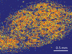
A novel machine-learning method to distinguish between tumor and normal tissue in optical coherence tomography (OCT) has been developed. Pre-clinical murine ear model implanted with mouse colon carcinoma CT-26 was used. Structural-image-based feature sets were defined for each pixel and machine learning classifiers were trained using “ground truth” OCT images manually segmented by comparison with histology. The accuracy of the OCT tumor segmentation method was then quantified by comparing with fluorescence imaging of tumors expressing genetically encoded fluorescent protein KillerRed that clearly delineates tumor borders. Because the resultant 3D tumor/normal structural maps are inherently co-registered with OCT derived maps of tissue microvasculature, the latter can be color coded as belonging to either tumor or normal tissue. Applications to radiomics-based multimodal OCT analysis are envisioned.
REFERENCES
- 1P.Lambin, E.Rios-Velazquez, R.Leijenaar, S.Carvalho, R. G. van Stiphout, P.Granton, C. M.Zegers, R.Gillies, R.Boellard, A.Dekker, Eur. J. Cancer 2012, 48, 441.
- 2C.Parmar, R. T.Leijenaar, P.Grossmann, E. R.Velazquez, J.Bussink, D.Rietveld, M. M.Rietbergen, B.Haibe-Kains, P.Lambin, H. J.Aerts, Sci. Rep. 2015, 5, 11044.
- 3E. R.Velazquez, C.Parmar, M.Jermoumi, R. H.Mak, A. van Baardwijk, F. M.Fennessy, J. H.Lewis, D.De Ruysscher, R.Kikinis, P.Lambin, Sci. Rep. 2013, 3, 3529.
- 4T. P.Coroller, P.Grossmann, Y.Hou, E. R.Velazquez, R. T.Leijenaar, G.Hermann, P.Lambin, B.Haibe-Kains, R. H.Mak, H. J.Aerts, Radiother. Oncol. 2015, 114, 345.
- 5D. A.Gutman, W. D.Dunn Jr.., P.Grossmann, L. A.Cooper, C. A.Holder, K. L.Ligon, B. M.Alexander, H. J.Aerts, Neuroradiology 2015, 57, 1227.
- 6A. F.Fercher, C. K.Hitzenberger, G.Kamp, S. Y.El-Zaiat, Opt. Commun. 1995, 117, 43.
- 7D.Huang, E. A.Swanson, C. P.Lin, J. S.Schuman, W. G.Stinson, W.Chang, M. R.Hee, T.Flotte, K.Gregory, C. A.Puliafito, Science 1991, 254, 1178.
- 8D. J.Faber, F. J.Van der Meer, M. C.Aalders, T. G. van Leeuwen, Opt. Express 2004, 12, 4353.
- 9M.Pircher, E.Goetzinger, R.Leitgeb, C.Hitzenberger, Opt. Express 2004, 12, 3236.
- 10B. F.Kennedy, K. M.Kennedy, D. D.Sampson, IEEE J. Sel. Top. Quant. Electron. 2014, 20, 272.
- 11V. Y.Zaitsev, I.Vitkin, L.Matveev, V.Gelikonov, A.Matveyev, G.Gelikonov, Radiophys. Quant. Electron. 2014, 57, 210.
- 12Z.Chen, T. E.Milner, D.Dave, J. S.Nelson, Opt. Lett. 1997, 22, 64.
- 13A.Mariampillai, B. A.Standish, E. H.Moriyama, M.Khurana, N. R.Munce, M. K.Leung, J.Jiang, A.Cable, B. C.Wilson, I. A.Vitkin, Opt. Lett. 2008, 33, 1530.
- 14L. A.Matveev, V. Y.Zaitsev, G. V.Gelikonov, A. L.Matveyev, A. A.Moiseev, S. Y.Ksenofontov, V. M.Gelikonov, M. A.Sirotkina, N. D.Gladkova, V.Demidov, Opt. Lett. 2015, 40, 1472.
- 15R. K.Wang, S. L.Jacques, Z.Ma, S.Hurst, S. R.Hanson, A.Gruber, Opt. Express 2007, 15, 4083.
- 16L.Conroy, R. S.DaCosta, I. A.Vitkin, Opt. Lett. 2012, 37, 3180.
- 17R.Reif, J.Qin, L.An, Z.Zhi, S.Dziennis, R.Wang, J. Biomed. Imag. 2012, 2012, 9.
- 18D.Sheet, A.Chaudhary, S. P. K.Karri, D.Das, A.Katouzian, P.Banerjee, N.Navab, J.Chatterjee, A. K.Ray, J. Biomed. Opt. 2013, 18.
- 19K.Vermeer, J.Van der Schoot, H.Lemij, J.De Boer, Biomed. Opt. Express 2011, 2, 1743.
- 20Y.Gan, D.Tsay, S. B.Amir, C. C.Marboe, C. P.Hendon, J. Biomed. Opt. 2016, 21.
- 21N.Gladkova, М.Sirotkina, N.Buyanova, Т.Kalganova, М.Karabut, V.Elagin, S.Kuznetsov, L.Snopova, G. Gelikonov, V. Y. Zaitsev. Sovremennye Tehnologii v Medicine. 2015, 7.
- 22Y.Jung, S.Dziennis, Z.Zhi, R.Reif, Y.Zheng, R. K.Wang, PLoS One 2013, 8, e57976.
- 23G. H.Dunteman, Principal Components Analysis, Sage, Newbury Park, CA, 1989.
10.4135/9781412985475 Google Scholar
- 24L.Breiman, Mach. Learn. 2001, 45, 5.
- 25J. A.Hanley, B. J.McNeil, Radiology 1982, 143, 29.
- 26J. A.Hartigan, J. R. Stat. Soc. Ser. C. Appl. Stat. 1979, 28, 100.
10.2307/2346830 Google Scholar
Citing Literature