Intrarenal pressure monitoring via flexible and navigable suction ureteral access sheath in retrograde intrarenal surgery: A preclinical animal study and a pilot clinical study
Wei Zhu, Steffi Kar Kei Yuen, Jianwei Cao, Chu Ann Chai, and Shusheng Liu contributed equally to this work.
Abstract
Background
Elevated intrarenal pressure (IRP) during retrograde intrarenal surgery (RIRS) can lead to deleterious complications. Emerging non-invasive, real-time IRP monitoring tools are proving crucial for enhancing procedural safety. This study evaluates a newly developed flexible and navigable suction ureteral access sheath (FANS) with IRP monitoring capabilities through animal study and a clinical trial, assessing its accuracy and operational benefits.
Methods
A preclinical animal study and a prospective clinical trial involving 100 patients were conducted. The animal study confirmed the accuracy of IRP-monitoring FANS, whilst the clinical trial compared its performance to conventional FANS in RIRS. The evaluated outcomes included the accuracy of IRP measurements, the irrigation flow rate, the duration of the operation, and the stone-free rate (SFR). Statistical comparisons were performed using appropriate tests with a significant threshold of p < .05. Registration for this study is recorded under the identifier NCT06729801 at ClinicalTrials.gov.
Results
In the animal study, IRP-monitoring FANS demonstrated high accuracy in real-time IRP measurement, comparable to percutaneous nephrostomy-based monitoring. In the clinical study, IRP-monitoring FANS enabled increased irrigation flow whilst maintaining safe IRP levels within 30 mmHg. Operative time was significantly shortened in IRP-monitoring FANS group (50.9 vs. 67.6 min, p < .01), with similar SFRs between groups. No notable discrepancies in the rates of complications were observed.
Conclusions
The IRP-monitoring FANS improves stone retrieval efficiency and shortens operative time whilst ensuring safety through real-time IRP monitoring. This novel device marks a major improvement in both the safety and effectiveness of RIRS for managing large renal stones.
1 INTRODUCTION
Recent advancements in endourology have enabled urologists to explore the boundaries of retrograde intrarenal surgery (RIRS) in the treatment of urolithiasis.1 Following the introduction of innovative laser systems and flexible and navigable suction ureteral access sheath (FANS),2 the scope of indications for RIRS has broadened, and the success rates of achieving a stone-free status have significantly increased.3, 4
The primary limitation of RIRS at present is its relatively low stone retrieval efficiency. Consequently, procedures for larger stone volume require extended operative times, which is a well-known factor for sepsis.5 Improving the stone retrieval efficiency of RIRS has thus become a major focus of recent research.4
In RIRS procedures using FANS, adequate irrigation helps to enhance suction and stone retrieval efficiency. However, increased irrigation during the procedure may lead to elevated intrarenal pressure (IRP), thereby raising the risk of postoperative infection. Thus, maintaining adequate irrigation and improving stone retrieval efficiency whilst preventing excessive IRP is crucial, making real-time monitoring of IRP a timely essential tool during the procedure.6, 7
We now report a type of FANS designed to monitor real-time IRP. A preclinical animal study has demonstrated that the IRP-monitoring FANS can accurately monitor intraoperative real-time IRP. A pilot clinical study has shown that using the IRP-monitoring FANS allows for real-time IRP monitoring, enabling surgeons to significantly increase irrigation whilst maintaining IRP within a safe range, enhancing suction and stone retrieval efficiency, and substantially shorten operative time. As a result, RIRS can be performed safely and effectively even when managing large renal stones.
2 METHODS
2.1 Device
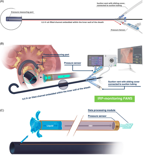
We utilised a novel type of FANS that is capable of real-time IRP monitoring (YiGao Med, China). The sheath's basic structure resembles that of a conventional FANS and is available in three lengths: 50 cm/55 cm were used for male patients, whilst 40 cm was designated for female patients. Sheath comes in two different sizes: 12/14 Fr and 10/12 Fr (inner diameter/outer diameter). Three millimetres from the distal tip of the sheath, a pressure-measuring port is connected to a 0.5 Fr air filled channel embedded within the inner wall of the sheath, which is connected to a pressure sensor (YiGao Med, China) at the external end. This embedded channel maintains the sheath's flexibility and outer diameter without altering the external physical characteristics of the sheaths (Figure 1a,b). When positioned in the kidney, pressure changes within the renal calyx are transmitted through the port, via the tiny embedded channel to the sensor in the external handle (Figure 1c). The real-time IRP is displayed on a monitor, allowing the surgeon to observe intraoperative IRP. This setup enables continuous IRP monitoring, capturing pressure variations at a rate of 5 measurements per second.

2.2 Preclinical animal study
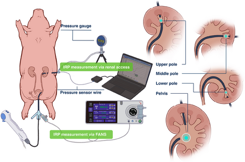
We utilised an in vivo adult porcine model to evaluate the accuracy of IRP monitoring by the IRP-monitoring FANS. After anaesthetising three adult female pigs, we created percutaneous nephrostomy tracts in the upper, middle and lower calyces using ultrasonographic guidance, and then dilated these to 14 Fr. A pressure sensor was then installed through the nephrostomy tract and connected to an external computer to display real-time IRP. Concurrently, a 12/14 Fr IRP-monitoring FANS was introduced into the ureter and advanced to the renal pelvis. Guided by a flexible ureteroscope, the IRP-monitoring FANS was sequentially positioned at various sites (e.g., upper, middle, lower calyx, and renal pelvis), with irrigation flow rates set at 50–100 mL/min and suction pressures set at 80 mmHg. The IRP values recorded by the FANS were compared with those obtained through the nephrostomy tract. Each site was measured for 10 min, with averages calculated to assess differences between sheath-measured and nephrostomy-measured IRP values (Figure 2).

2.3 Pilot clinical study
From January to December 2024, a prospective, comparative cohort study was carried out at a tertiary medical center. Two experienced surgeons, each with over 1000 RIRS procedures, performed all surgeries. The study included consecutive patients undergoing RIRS for renal stones, utilising either conventional FANS or the IRP-monitoring FANS. Participants met the following criteria: (1) adults with renal stones sized between 1–4 cm, and (2) preoperative intravenous pyelography (IVP) showing no significant ureteral stricture. Registration details for this research can be found at ClinicalTrials.gov under the identifier NCT06729801.
Standard preoperative protocols followed those outlined in our earlier study.3 All participants underwent a non-contrast computed tomography (CT) scan and IVP prior to surgery to measure stone size and density uniformly using the same software.8 Patients with negative urine cultures received standard perioperative antibiotic prophylaxis administered 30 min before the procedure. For those with positive preoperative urine cultures, appropriate antibiotics were prescribed based on culture sensitivity results and administered for 4–7 days prior to undergoing RIRS.
Our approach to RIRS extensively detailed in a prior publication3 and is summarised herein. The patient was placed in the lithotomy position, and the procedure was conducted under general anaesthesia. A guidewire was advanced into the renal pelvis. Either a 12/14 Fr FANS or IRP-monitoring FANS was used (YiGao Med, China). If the sheath could not be inserted, a smaller 10/12 Fr sheath was employed as an alternative.
A 7.5 Fr disposable flexible ureteroscope manufactured by Pusen, China was employed during the RIRS procedures. For patients utilising the conventional FANS, intraoperative irrigation was performed with an irrigation pump set at a flow rate of 40–80 mL/min, whilst negative pressure suction was maintained at 80 mmHg. In the IRP-monitoring FANS group, the initial irrigation flow rate was also set at 40–80 mL/min; however, the surgeon could gradually increase the flow rate based on the real-time IRP values, whilst ensuring that the IRP remained within 30 mmHg. The maximum irrigation flow rate could be increased up to 200 mL/min, whilst the negative pressure suction ranged from 80 to 120 mmHg. Stone fragmentation was performed using a holmium laser with a 200 µm fibre, setting the energy below 30 W. Fragments small enough to navigate through the space between the ureteroscope and the sheath were suctioned out, and larger fragments were manually extracted by retracting the flexible ureteroscope. Following each procedure, a 6 Fr JJ stent was inserted, remaining in place for 1–2 weeks. The regular postoperative placement of a Foley catheter was not practised.
All patients underwent a low-dose CT scan on the morning following the procedure. Stone-free status was determined by the absence of residual stones or fragments bigger than 2 mm as identified on the CT scans.
The primary outcome of the pilot study was operating time, which was defined as the time when the flexible ureteroscope was inserted per urethra until the time of stent placement upon completion. Secondary outcomes include stone-free rate (SFR), the duration of postoperative hospitalisation, and complications, which were evaluated using the modified Clavien-Dindo classification.9
2.4 Statistical analysis
The statistical comparisons between the two groups were carried out using the t-test, Pearson's Chi-square test and Fisher's exact test. Statistical significance was defined as a p-value below.05. Data analysis was performed using SPSS software, version 18.0 (IBM Corp., Armonk, NY, USA).
3 RESULTS
3.1 Preclinical animal study
Our animal study showed that the IRP measurements taken via IRP-monitoring FANS were consistent in accuracy with those taken through the percutaneous renal access. The difference in range between the two methods was 0.011 mmHg (95% confidence interval (CI) −0.004 to 0.026, p = .14). Moreover, IRP measured through FANS were consistent with those measured via percutaneous renal access, regardless of whether they were taken in the renal pelvis, upper calyx, middle calyx or lower calyx (Table 1). In conclusion, the animal study results suggested that IRP monitoring FANS can accurately reflect the real-time IRP.
| IRP-monitoring FANS | Renal access | Mean difference (95% CI) | p-Value | |
|---|---|---|---|---|
| Mean IRP (mmHg) | 10.006 (0.585) | 9.994 (0.578) | 0.011 (−0.004 to 0.026) | .14 |
| Location | ||||
| Upper calyx | 10.009 (0.591) | 9.990 (0.577) | 0.019 (−0.011 to 0.048) | .21 |
| Middle calyx | 10.009 (0.580) | 9.993 (0.579) | 0.016 (−0.014 to 0.045) | .29 |
| Lower calyx | 9.998 (0.584) | 9.994 (0.580) | 0.004 (−0.025 to 0.034) | .78 |
| Renal pelvic | 10.006 (0.585) | 10.000 (0.578) | 0.006 (−0.023 to 0.036) | .69 |
- Note: Data are presented as mean (standard deviation).
- Abbreviations: CI, confidence Interval, IRP, Intrarenal pressure; FANS, flexible and navigable suction ureteral access sheath.
3.2 Pilot clinical study
One hundred patients who received RIRS using either conventional FANS or the IRP-monitoring FANS participated in the study. Baseline characteristics for both patient groups are detailed in Table 2. No significant differences were observed between the groups in terms of age, body mass index (BMI), stone burden, comorbidities, hydronephrosis severity and positive preoperative urine cultures.
| Conventional FANS (n = 50) | IRP-monitoring FANS (n = 50) | p-value | |
|---|---|---|---|
| Preoperative clinical characteristics | |||
| Age (yr) | 52.1 (14.2) | 51.9 (10.8) | .94 |
| Gender, n (%) | |||
| Male | 30 (60.0) | 28 (56.0) | .69 |
| Female | 20 (40.0) | 22 (44.0) | |
| BMI (kg/m2) | 23.8 (4.7) | 22.5 (4.3) | .15 |
| ASA classification, n (%) | |||
| I | 18 (36.0) | 23 (46.0) | .81 |
| II | 32 (64.0) | 27 (54.0) | |
| Laterality, n (%) | |||
| Left | 24 (48.0) | 25 (50.0) | .84 |
| Right | 26 (52.0) | 25 (50.0) | |
| Stone diameter (mm) | 22.0 (3.1) | 23.3 (4.4) | .09 |
| Stone surface (mm2) | 339.0 (118.5) | 374.5 (107.3) | .12 |
| Stone type, n (%) | |||
| Single | 34 (68.0) | 28 (56.0) | .21 |
| Multiple | 16 (32.0) | 22 (44.0) | |
| CT value of stone (HU) | 1166 (312) | 1079 (381) | .21 |
| Grade of hydronephrosis, n (%) | |||
| None or mild | 26 (52.0) | 29 (58.0) | .55 |
| Moderate or severe | 24 (48.0) | 21 (42.0) | |
| Comorbidities, n (%) | |||
| Hypertension | 14 (28.0) | 16 (32.0) | .66 |
| Diabetes | 2 (4.0) | 3 (6.0) | .65 |
| Initial positive urine culture, n (%) | 10 (20.0) | 13 (26.0) | .48 |
| Pre-stenting, n (%) | 2 (4.0) | 4 (8.0) | .40 |
| Operative characteristics | |||
| Sheath size (Fr), n (%) | |||
| 12/14 | 32 (64.0) | 35 (70.0) | .52 |
| 10/12 | 18 (36.0) | 15 (30.0) | |
| Irrigation fluid volume (mL/min) | 59.6 (17.2) | 119.8 (27.4) | <.01 |
| Baseline IRP (mmHg) | N/A | 5.4 ± 2.6 | |
| Intraoperative IRP (mmHg) | N/A | 13.8 ± 4.7 | |
| Number of patients who had one episode of IRP ≥ 30 mmHg | N/A | 6 (12.0) | |
| Operative outcomes | |||
| Operative time (min) | 67.6 (23.5) | 48.7 (17.2) | <.01 |
| Immediately SFR, n (%) | 44 (88.0) | 46 (92.0) | .51 |
| 3 months SFR, n (%) | 47 (94.0) | 48 (96.0) | 1 |
| Use of stone basket, n (%) | 5 (10.0) | 2 (4.0) | .44 |
| Post-operative hospital stays (days) | 1.4 (0.7) | 1.3 (0.4) | .38 |
| Complications, n (%) | |||
| Fever (> 38.5°C) Clavien grade I | 3 (6.0) | 1 (2.0) | .62 |
- Note: Data are presented as mean (standard deviation), or number (proportion).
- Abbreviations: IRP, Intrarenal pressure; FANS, flexible and navigable suction ureteral access sheath.
No notable statistical difference was found in the sheath sizes used between the IRP-monitoring FANS group and the conventional FANS group. Seventy percent of patients in the IRP-monitoring FANS group and 64% of patients in the conventional FANS group used 12/14 Fr sheaths (p = .52) (Table 2).
IRP was measured in all cases in the IRP-monitoring FANS group. The overall IRP in the IRP-monitoring FANS group was 13.8 ± 4.7 mmHg. Twelve percent of patients in the IRP-monitoring FANS group had one episode of IRP ≥ 30 mmHg. However, the duration was very short, with the cumulative time exceeding IRP ≥ 30 mmHg being no more than 5 s. In the IRP-monitoring FANS group, the surgeon can appropriately increase or adjust intraoperative irrigation based on real-time IRP values to improve the efficiency of stone extraction by suction during the procedure. The IRP-monitoring FANS group exhibited a higher irrigation flow rate during RIRS when compared to the conventional FANS group (119.8 ± 27.4 vs. 59.6 ± 17.2 mL/min, p < .01). The IRP-monitoring FANS had a shorter operative time than the conventional FANS group (48.7 vs. 67.6 min, p < .01). The SFRs were similar between conventional and IRP-monitoring FANS respectively (immediately SFR 88% vs. 92%, p = .51; overall SFR at 3 months post-operatively 94% vs. 96%, p = 1.00) (Table 2).
The overall rates of operative complications, as evaluated using the modified Clavien–Dindo grading system, were comparable across both groups. No differences were observed in the incidence of postoperative fever, and neither group experienced cases of urosepsis. Additionally, the duration of postoperative hospitalisation was not significantly different between the two groups (Table 2).
To further explore whether the size of the sheath affects irrigation flow rate, SFRs and IRP, we performed a subgroup analysis of the IRP-monitoring FANS group based on the sheath size. The results showed no significant statistical difference in irrigation flow rate and SFRs between the 12/14 Fr and 10/12 Fr sheaths. The intraoperative IRP was slightly higher in the 10/12 Fr group compared to the 12/14 Fr group (13.0 ± 5.0 vs. 15.6 ± 3.5 mmHg, p = .08), but still within the safe IRP range (≤30 mmHg) (Table S1).
4 DISCUSSION
The advent of FANS is a milestone in the advancement of RIRS.10 However, uncontrolled IRP during surgery, along with suboptimal irrigation and suction settings, remains critical challenges in RIRS procedures. The introduction of IRP-monitoring FANS offers an effective tool in addressing these issues whilst enhancing both surgical safety and efficiency.
This study demonstrates that the IRP-monitoring FANS provides accurate real-time IRP measurements during RIRS, enabling surgeons to safely increase intraoperative irrigation. This approach not only improves suction and stone retrieval efficiency but also significantly shortens operative time, enabling RIRS to become a practical and effective option for managing larger stones (Figure 3). To our knowledge, this study is the first to effectively implement real-time IRP monitoring during RIRS with the use of a FANS.

Currently, the primary method for monitoring IRP during RIRS relies on pressure-sensing ureteroscopes for the real-time measurement.6, 11 These ureteroscopes are equipped with a pressure sensor located at their distal tip. However, there are several limitations to using flexible ureteroscopes for IRP monitoring. First, IRP-monitoring flexible ureteroscopes are relatively expensive due to the specialised miniaturised pressure sensor technology incorporated at its tip, significantly increasing production costs. Consequently, these scopes are often much costlier than standard flexible ureteroscopes, making them less accessible, particularly in developing regions where cost-sensitive urologists are unlikely to adopt them solely for the IRP-monitoring feature. Second, the engineering of the pressure sensor into the flexible ureteroscope necessitates a larger scope size (8.6–9.5 Fr)12 limiting the development of smaller, more versatile designs. Third, the distal pressure sensor is prone to damage during laser lithotripsy, which can result in measurement failure. Finally, when using a FANS, the flexible ureteroscope is frequently withdrawn to the sheath's external end to facilitate fragment extraction. During this process, the displayed pressure no longer reflects real-time IRP within the kidney, limiting the accuracy of monitoring.
Compared to pressure-sensing flexible ureteroscopes, IRP-monitoring FANS offer several unique advantages. First, they utilise air conduction for pressure detection, making them significantly more cost-effective than pressure-sensing ureteroscopes, with a price comparable to that of conventional FANS. Additionally, the air filled channel is exceptionally thin—only 0.5 Fr—and embedded within the sheath wall, ensuring that it does not affect the sheath's diameter or flexibility. The pressure measurement port is strategically positioned 3 mm from the sheath's tip, minimising the risk of damage during laser lithotripsy. Furthermore, because the sheath remains in the collecting system throughout the procedure, it allows for continuous, real-time IRP monitoring within the pelvicalyceal system, provided it is properly positioned. However, IRP-monitoring FANS also have certain limitations. In some cases, blood clots or stone dust may block the pressure port on the sheath, potentially resulting in inaccurate IRP measurements.
This study is subject to several limitations. First, the relatively small-sample size of the clinical trial may restrict the broader applicability of the results. Second, further validation of the benefits of IRP-monitoring FANS in RIRS will require additional randomised controlled trials.
5 CONCLUSION
The IRP-monitoring FANS accurately reflects the real-time IRP during RIRS, enabling surgeons to safely increase intraoperative irrigation to enhance the efficiency of stone fragmentation, suction and extraction. This ensures procedural safety whilst significantly improving surgical outcomes. As a result, the IRP-monitoring FANS represent a major technological advancement, improving stone extraction efficiency, potentially broadening the indications for RIRS.
AUTHOR CONTRIBUTIONS
Guohua Zeng and Wei Zhu conceived and designed the study. Wei Zhu, Jianwei Cao, Shusheng Liu, Wen Zhong and Yongda Liu acquired the data. Wei Zhu, Shusheng Liu, Jingzeng Du and Guohua Zeng analysed and interpreted the data. Wei Zhu and Chu Ann Chai drafted the manuscript. Steffi Kar Kei Yuen critically revised the manuscript for important intellectual content. Wei Zhu and Shusheng Liu conducted the statistical analysis. Wei Zhu and Guohua Zeng obtained funding. Wei Zhu and Jianwei Cao provided administrative, technical, or material support. Guohua Zeng supervised the study. All authors have read and agreed to the published version of the manuscript.
ACKNOWLEDGMENTS
The authors would like to express their gratitude to Tao Huang for his assistance with the illustrations in this paper. This work was financed by grants from Plan on enhancing scientific research in Guangzhou Medical University [Grant number: GMUCR2024-01006] and Natural Science Foundation of Guangdong Province [Grant number: 2024A1515011644].
CONFLICT OF INTEREST STATEMENT
On behalf of all authors, Guohua Zeng declared a potential conflict of interest, as YiGao Medical Technology Co., Ltd, the manufacturer of the IRP-monitoring FANS and system, provided IRP-monitoring FANS to centre as an in-kind contribution. No other payments were made to the institutions, hospitals, or any of the authors related to this study. The only support provided by the company was the in-kind contribution of the IRP-monitoring FANS.
ETHICS STATEMENT
This study was approved by the Institutional Review Board of The First Affiliated Hospital of Guangzhou Medical University [EC-2023-037(XJS)]. Written informed consent was obtained from all patients. All procedures were in accordance with the Declaration of Helsinki.
Open Research
DATA AVAILABILITY STATEMENT
Data are not available for sharing due to privacy and ethical or legal issues. Summary statistical data will be available from the corresponding author on reasonable request.


