Human epidermal growth factor receptor 2 status in urothelial bladder carcinoma: Frequency, genomic patterns and clinical significance
[Correction added on 30 November 2023; after first online publication missing sub-sections have been added.]
Tianxi Yu Huiying Tao Wei Lv Ning Liang contributed equally.
Dear Editor,
Urothelial bladder carcinoma (UBC), represents a formidable global public health concern, accounting for about 80% of UC cases.1 Therapeutic options include transurethral resection of bladder tumors and bladder perfusion chemotherapy for early-stage UBC. However, locally advanced unresectable or metastatic UC (LA/mUC) frequently presents therapeutic obstacles with limited efficacy.2 Human epidermal growth factor receptor 2 (HER2), has been shown associated with prognosis in breast and gastric cancer. However, the HER2 status in UBC is not fully understood, and a standardized method for its detection is yet to be established.
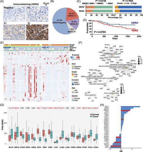
This study included 79 UBC patients who underwent surgical treatment at Yantai Yuhuangding Hospital from 2017 to 2021. Two patients received chemotherapy before surgery, while the rest of the patients did not receive chemotherapy or radiation therapy before surgery. The corresponding clinical data were collected (Table S1). The tumour samples were collected for immunohistochemistry and RNA sequencing. The HER2 immunohistochemical results were scored based on the American Society of Clinical Oncology HER2 testing guidelines (2018).3 The scoring system used was: A score of 3 indicated strong membrane staining in over 10% of cells; a score of 2 represented weak to moderate complete membrane staining in over 10% of cells; a score of 1 indicated faint and partial membrane staining in over 10% of cells, while a score of 0 denoted either unstained tumour cells or less than 10% staining. An immunohistochemistry (IHC) score of 3+ or 2+ signified HER2 high expression or overexpression, while a score of 0 or 1+ indicated HER2 low expression (Figure 1A). Among the 79 CCGA-UBC patients included in this study, 45 (57%) had an IHC score of 0, 16 (20.3%) had a score of 1+, 12 (15.2%) had a score of 2+ and six (7.6%) had a score of 3+ (Figure 1B). Overall, 18 cases (22.8%) were found to have a high HER2 expression (IHC 2+/3+). It is worth noting that the incidence of HER2 overexpression in UBC ranges from 4%-76% in different literature, which may be attributed to variations in tissue sample sources.4-6

Among the 25 patients who received postoperative chemotherapy or radiation therapy, 10 showed HER2 overexpression, suggesting a possible association between HER2 overexpression and improved survival and increased HER2 prevalence. During follow-up, 30% of patients with HER2-positive died. Among these 25 patients, 9 died with a median overall survival time of 630 days. The remaining 16 patients had a median progression-free survival time of 1317 days. There was no significant difference in tumour stage and grade between HER2-negative and positive patients (Fisher's test; p > .05) (Figure 1C). HER2-positive patients exhibited a more favorable prognosis compared to HER2-negative patients. However, due to the limited sample size, there is no clear association between prognosis and HER2 expression (log-rank test; p = .0793) (Figure 1D). Previous studies investigating genetic variants related to UBC have yielded conflicting conclusions regarding the prognostic value of HER2 status.7 While some research suggests that HER2 upregulation is linked to poor prognosis, others find no effect on prognosis.8
We next explored the mRNA aberrations associated with HER2 expression in the clinical cohort. Transcriptome data analysis of 70 patients revealed 312 differentially expressed genes (DEGs). Among the DEGs, the top five genes were MYB, CTSG, HSPB7, SYNPO2 and CNN1 (Figure 1E). Pathway enrichment analysis of the DEGs indicated involvement in pathways such as muscle system process, muscle contraction, extracellular structure organization, and cytokine−cytokine receptor interaction (Figure 1F). This prompts the hypothesis that HER2 might play a role within these signaling pathways, potentially linking it to UBC.
Finally, we conducted a pan-cancer analysis using the TCGA database to explore the differences in HER2 expression between tumors and adjacent tissues. We observed significant variations in HER2 expression among several cancer types, such as UBC, Breast invasive carcinoma, and so on (p < .01). However, there was no clear difference in the expression of HER2 in tumors such as colon adenocarcinoma (COAD), esophageal carcinoma (ESCA), head and neck squamous cell carcinoma (HNSC), and so on (p > .01) (Figure 1G). Furthermore, we evaluated the correlation between HER2 expression and prognosis across multiple cancer types. The findings suggest that HER2 detection may not be adequate for identifying a patient subgroup with an unfavorable prognosis across various cancer types, with the exception of mesothelioma (MESO), skin cutaneous melanoma (SKCM), ovarian serous cystadenocarcinoma (OV) and brain lower grade glioma (LGG) (Figure 1H).
CONCLUSION
This study offers a valuable methodology and reference for evaluating HER2 status at UBC. However, to validate these findings, further prospective studies involving larger sample sizes and multiple detection methods are needed. In conclusion, our study contributes to the understanding of HER2 expression in UBC and lays the foundation for establishing a standardized approach to assess HER2 expression. The insights gained from our results are of significant importance for future investigations and may guide the development of novel therapeutic strategies for individuals with UBC.
AUTHOR CONTRIBUTIONS
Data analysis, H.T., W.L., and N.L.; Funding acquisition, C.L.; Methodology, W.L., C.L., and G.Y.; Supervision, W.W., C.L., and G.Y.; Visualization, H.T., W.L., and T.Y.; Information collection, T.Y., X.C., N.L., and S.W.; HER2 staining, N.L., X.C., and Y.L.; Writingoriginal draft, T.Y., and W.L.; All authors have contributed to the execution of the experiments and studies.
ACKNOWLEDGMENTS
We thank all the patients for their participation in the study. W.L. was funded by the China Scholarship Council (CSC).
CONFLICT OF INTEREST STATEMENT
The authors declare no conflict of interest.
FUNDING INFORMATION
This research was funded by the Taishan Scholar Program (No. Tsqn202103198) to Chunhua Lin.
ETHICS APPROVAL
This study has obtained ethical approval from the IRB of Yantai Yuhuangding Hospital.
Open Research
DATA AVAILABILITY STATEMENT
All sequencing data can be acquired from GSA-Human with accession number HRA003461.


