Slipped capital femoral epiphysis in an adolescent with congenital adrenal hyperplasia: A case report
Key Clinical Message
In previous reports, hypothyroidism, hypopituitrism, and hypogonadism were common endocrine causes of SCFE, but this is the first time that congenital adrenal hyperplasia has been observed. As such, patients who have undergone long-term endocrine treatment for congenital adrenal hyperplasia could potentially be subjected to a higher risk for SCFE.
1 INTRODUCTION
Slipped capital femoral epiphysis (SCFE) is a complication of the proximal femur commonly seen in adolescents. It is defined as the displacement of the metaphysis relative to the epiphyses1 A previous study regarding SCFE in the Asian population indicated that the incidence in children 9 to 14 years of age was 6.0/100,000,2 with obesity and puberty being important risk factors.3 In recent years, endocrine disorders such as hypothyroidism, hyperparathyroidism, hypogonadism, and hypopituitarism have increasingly been reported as causes of SCFE.4 Congenital adrenal hyperplasia (CAH) is a group of autosomal recessive disorders that includes enzyme deficiencies in the adrenal steroidogenesis pathway, which results in impaired cortisol biosynthesis.5 To the best of our knowledge, this is the first report of SCFE in a 12-year-old boy who was also diagnosed with CAH.
2 CASE HISTORY/EXAMINATION
The timeline for this case report is shown in Figure 1. A 12-year-old Chinese boy suddenly felt severe pain in his right hip as he was walking on the level ground at school and was subsequently referred to our hospital. The pain started to appear 2 weeks before his hospitalization with no external trauma. A comprehensive physical examination was performed according to SCFE and CAH. The patient's right lower limb showed external rotation and short deformity. His right hip passive range of motion was as follows: extension 0°; flexion 60°; adduction 10°, and abduction 20°; severely restricted internal and external rotation. His muscle strength was 5/5, but he could not ambulate even with crutches. Combined with his symptoms, physical examination, and imaging examination, the patient was diagnosed with acute unstable SCFE according to the Loder and Fahey criteria.6
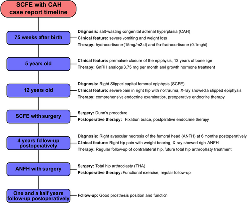
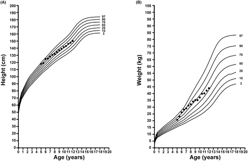
When this patient was 75 weeks old after birth, he was diagnosed with CAH on account of severe vomiting and weight loss at department of endocrinology of our hospital. According to the CAH classification, the patient in this case belongs to the salt-wasting type, which is characterized by glucocorticoid (GC) deficiency, mineralocorticoid deficiency, and accumulation of sex hormones. He was subsequently treated with hydrocortisone (15 mg/m2.d) and 9α-fludrocortisone (0.1 mg/d). The excess androgen presence led the child to have premature growth of pubic hair, rapid skeletal growth, and premature epiphyseal fusion. At the age of 5 years old, his bone age was tested to be already 13. It can be seen from the height growth chart (Figure 2) that at 5 years old, the patient had a height advantage over that of his peers, but as he got older, the advantage diminished. Under his parents' desire to improve their child's height, the patient started receiving gonadotropin-releasing hormone (GnRH) analogs under the guidance of an endocrinologist. The initial dosage was 0.82 mg per month and increased to 3.75 mg. This was taken in conjunction with growth hormone treatment. The patient's height increased with an average increase of 5–6 cm per year, the growth velocity has dropped to 4 cm per year in the past 2 years. Under the antiandrogenic effect of gonadotropin-releasing hormone analogs, the bone age of the child did not increase significantly. At the time of admission, the patient was 150 cm tall, weighed 43.8 kg, and had a normal body mass index (BMI) of 19.5 kg/m2. Due to CAH-induced precocious puberty, the patient had dark and dry skin; his Tanner stage was V for genital and pubic hair development. The patient's overall medical history led us to think about whether there was a link between endocrine disorders caused by CAH and SCFE.
3 METHODS
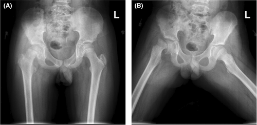
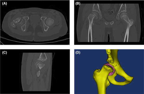
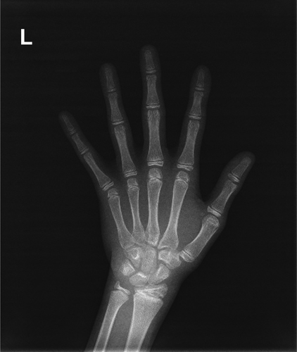
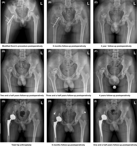
An anteroposterior pelvic radiograph (Figure 3) showed severe SCFE in the right hip. And a pelvic CT and three-dimensional reconstruction via the Mimics 19.0 software (Materialize, Leuven, Belgium) were completed (Figure 4), providing a more accurate position of the metaphyseal as well as a precise preoperative simulation of the length and position of the hollow screw. Radiological examinations of the right hip ruled out femoral fractures and tumors. An adrenal CT showed left adrenal hyperplasia (Figure 5), and an anteroposterior left-hand radiograph showed that his bone age was 13 years (Figure 6). Considering that the occurrence of SCFE in the patient might be related to his CAH history, a comprehensive endocrine examination was performed. Detailed laboratory testing results are shown in Table 1. Under the guidance of an endocrinologist, the patient's hydrocortisone was doubled before surgery and 2–3 days after. 40 mg was administered intravenously before the operation and once a day for 2 days after the operation. The GnRH analogs and growth hormone were suspended. The surgery improved the position of the femoral head relative to the femoral neck and was fixed using modified Dunn's procedure.7 (Figure 7). The operator performed a trochanteric flip osteotomy and a z-shaped capsulotomy anterior to the lesser trochanter through a posterolateral hip approach and an anterior dislocation of the hip. The femoral epiphysis is temporarily pinned with 2 mm-Kirschner wires. A 6.5 mm in diameter and 84 mm in length hollow compression screw was placed anterograde starting at the fovea of the femoral head, and a hollow lag screw and a Kirschner wire were used to fix the greater trochanter.

| Normal values | ||
|---|---|---|
| Cortisol (nmol/L) |
0 a.m.: 311.39 8 a.m.: 28.48 4 p.m.: 16.74 |
0 a.m.: 240–619 8 a.m.: 240–619 4 p.m.: <276 |
| ALD (ng/ml) | 0.13 | 0.06–0.174 |
| Serum Potassium (mmol/L) | 3.99 | 3.5–5.3 |
| Serum Sodium (mmol/L) | 136.5 | 137–147 |
| ACTH (pmol/L) |
0 a.m.: 5.97 8 a.m.: 440.40 4 p.m.: 53.59 |
0 a.m.: 1.6–13.9 8 a.m.: 1.6–13.9 4 p.m.: 1.6–13.9 |
| Progesterone (nmol/L) | 18.82 | 0.318–2.671 |
| Testosterone (nmol/L) | 0.84 | 6.07–27.1 |
| Estradiol (pmol/L) | 75 | <73.4–172.49 |
| FSH (mIU/ml) | 0.55 | 55.97–278.36 |
| LH (mIU/ml) | <0.20 | 1.24–8.62 |
| PRL (mIU/ml) | 347.31 | 55.97–278.36 |
| GH (ng/ml) | 0.658 | 0.003–0.971 |
| TSH (uIU/mL) | 4.45 | 0.27–4.2 |
| FT3 (pmol/L) | 7.20 | 3.1–6.8 |
| FT4 (pmol/L) | 17.93 | 12–22 |
4 RESULTS/OUTCOME AND FOLLOW-UP
A right hip abduction brace was used to immobilize the hip and make the right hip have moderate abduction (30 degrees). Partial weight bearing was performed 3 months postoperatively. The hydrocortisone and 9α-fludrocortisone resumed after the patient's diet returned to normal. Unfortunately, at the 6-month follow-up, the patient suffered from right avascular necrosis of the femoral head (ANFH). Four years after modified Dunn's procedure, the patient underwent right total hip arthroplasty (THA). So far, a follow-up of 1 and a half years has shown good prosthesis position and function. In addition, we paid close attention to the risk of contralateral epiphyseal slip during follow-up. At present, the contralateral epiphyseal tends to fusion, thus reducing the risk of slip.
5 DISCUSSION
SCFE usually occurs when there is a growth spurt during puberty, with the risk compounding in cases of obesity and abnormal anatomy of the proximal femur.8 In recent years, more and more attention has been given to the role of endocrine factors in the occurrence of SCFE. In previous reports, hypopituitarism, hypothyroidism, and hypogonadism were common endocrine causes of SCFE.9-12 CAH is also an endocrine disease characterized by hormonal disorders. The main hormones involved are adrenocortical hormones and sex hormones.5 This is the first report of SCFE in a child who was diagnosed with CAH.
The linear growth of childhood results from endochondral ossification in the growth plate. During adolescence, endocrine levels change greatly, and disturbances in endocrine mechanisms may lead to a weak physiological function of the proximal femur. Therefore, the effects of several hormones on the epiphyseal plate should be understood. Prepubertal growth is mainly regulated by the growth hormone-insulin-like growth factor 1 (GH-IGF-1) axis and the thyroid hormone (T3), while sex steroids are major regulators of pubertal growth. Increased sex hormones can also lead to increased GH-IGF-1 axis activity. GH can stimulate chondrocyte proliferation. IGF-1 is a key factor in ossification of the epiphyseal cartilage. Thyroid hormone promotes bone maturation and longitudinal growth, but excess thyroid hormone promotes early bone closure. A physiological dose of GC is beneficial for normal growth, but excess GC inhibits growth by inhibiting chondrocyte proliferation and cartilage matrix synthesis.13, 14 Previous studies have shown that growth hormone was safe for improving height.15 Nonetheless, SCFE seems to have spontaneously occurred in this patient with no clear cause. We speculate on two possible reasons. First, the epiphyseal plate was not closed due to the application of GnRH analogs, which increased the risk of SCFE. Second, the long-term use of GC could have affected chondrocyte proliferation and cartilage matrix synthesis, which caused unbalanced growth of the epiphyseal plate.
A study of adolescent SCFE by Julio Javier Masquijo16 showed that the incidence of ANFH after surgery was 47.6% (10/21 hips). As we all know, GC is a common cause of ANFH. This patient recovered from the use of GC after surgery due to CAH therapy. Thus, the possibility of ANFH may be higher when GC continues to be used after surgery. Therefore, we should be cautious about the occurrence of SCFE in the process of this treatment. In addition, comprehensive endocrine examination should be performed preoperatively to prevent adrenal crisis and imbalance of serum sodium and potassium, especially in children with salt-wasting type CAH.
6 CONCLUSION
In previous reports, hypothyroidism, hypopituitarism, and hypogonadism were common endocrine causes of SCFE, but this is the first time that congenital adrenal hyperplasia has been observed. As such, patients who have undergone long-term endocrine treatment for congenital adrenal hyperplasia could potentially be subjected to a higher risk for SCFE.
AUTHOR CONTRIBUTIONS
Yi-Fan Huang: Data curation; methodology; software; visualization; writing – original draft. Jin-Han Qiao: Methodology; software; visualization. Jia Zheng: Supervision; writing – review and editing.
ACKNOWLEDGMENTS
We would like to thank Cathy Lv for English language editing.
FUNDING INFORMATION
No funding provided.
CONFLICT OF INTEREST STATEMENT
All authors declare that they have no conflict of interest.
ETHICS STATEMENT
All procedures performed in the study were in accordance with the ethical standards of the ethics committee of Henan Provincial People's Hospital and national research committee and conformed to the 1964 Helsinki declaration.
CONSENT
Written informed consent was obtained from the patient's parents for publication of this case report and any accompanying images. A copy of the written consent is available for review by the Editor of this journal.
Open Research
DATA AVAILABILITY STATEMENT
The data that support the findings of this study are available from the corresponding author upon reasonable request.


