Teaching NeuroImages: Absence of bilateral internal carotid arteries with multiple aneurysms
Abstract
Absence of bilateral internal carotid arteries is a rare congenital vascular dysplasia. We reported the case and present some typical and exquisite neuroimges for teaching.
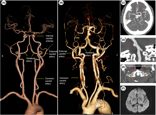
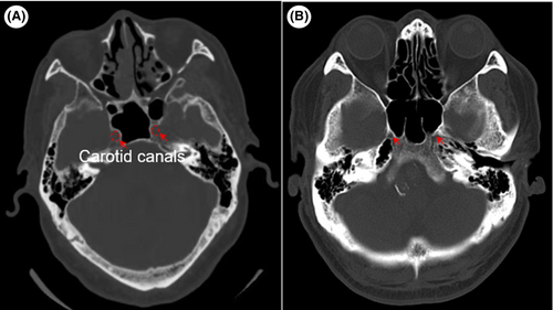
A 74-year-old male patient presented with aggravated weakness of both lower limbs for days. He was suspected diagnosis of cerebral ischemic disease and craniocervical CT angiography (CTA) were conducted. CTA revealed the absence of bilateral internal carotid arteries (ICAs) with compensated artery dilatation, multiple aneurysms, and sclerosis in cephalic and cervical arteries, without acute infarcts in diffusion-weighted images (DWI) (Figure 1). CT scan also revealed hypoplastic carotid canals at both sides in bone window (Figure 2). With the informed risk of aneurysm rupture, the patient insisted on being discharge after the limbs weakness was improved by symptomatic treatments for a week. Since then, no stroke happened during a year of follow-up.


The absence of internal carotid artery (ICA) is a rare congenital vascular dysplasia with an incidence rate less than 0.01%, mostly shown by unilateral left.1 The patients with the absence of ICA generally show developmental delay, subarachnoid hemorrhage, headache, dizziness, eye disease, and tinnitus with compensatory vessel-induced pulsive mass behind tympanum, basilar artery dilation, and abnormal ophthalmic artery originated from middle cerebral artery (MCA) or middle meningeal artery (MMA).2 Few patients with the absence of ICA are asymptomatic, in which the dilated vertebrobasilar artery and intact circle of Willis can be compensatory. The incidence of intracranial aneurysm associated with an increased hemodynamic load is 10-folds higher than that of the normal population.3 The current treatment with the absence of ICA is symptomatic or rehabilitative, and collateral circulation reconstruction and surgical or endovascular repair to reduce the risk of aneurysm rupture are also important interventions to improve the survival rate of patients.4
In conclusion, we present here teaching neuroimages for the absence of bilateral ICA and multiple intracranial aneurysm in an old patient. The results also indicated that the vertebrobasilar artery and the intact circle of Willis play an important role in the compensation of global cerebral blood flow. However, changes in cerebral hemodynamics may increase the risk of aneurysmal genesis.
AUTHOR CONTRIBUTIONS
Yang Gao: Data curation; funding acquisition; methodology; writing – original draft. jianhua yuan: Conceptualization; data curation; software; writing – original draft. jianzhi wang: Funding acquisition; project administration; writing – review and editing.
ACKNOWLEDGMENTS
The authors thank Dr. Dan Lin and the patient for permission to report this case.
FUNDING INFORMATION
Funded by the Wuhan Health Science Foundation (WX20Q04), the Fundamental Research Funds for the Central Universities (YCJJ202203019).
CONFLICT OF INTEREST
The authors declare that there are no conflict of interests.
CONSENT
Written informed consent was obtained from the patient to publish this report in accordance with the journal's patient consent policy.
Open Research
DATA AVAILABILITY STATEMENT
The data that support the findings of this study are available on request from the corresponding author. The data are not publicly available due to privacy or ethical restrictions.


