Bilateral non-contiguous necrotizing fasciitis of the lower extremities
Abstract
Necrotizing fasciitis (NF) is an uncommon soft tissue infection. Multifocal-extremity NF is a rarity with high mortality rates. Herein we report a case of bilateral non-contiguous NF of the lower extremities due to Escherichia coli with a fatal outcome, stressing the necessity of rapid and aggressive intervention in suspected cases.
1 INTRODUCTION
The term necrotizing fasciitis (NF) describes a group of rare, but potentially life-threatening invasive bacterial infections of the skin and subcutaneous soft tissues, which tend to progress fast along the fascia, causing fulminant tissue destruction. Pathomechanistically NF is characterized by synergistic effects of bacterial virulence factors (toxins, enzymes) and a disorder in the humoral immune defense of susceptible hosts, leading to intravascular thrombosis and ischemic necrosis.1 The incidence has been reported to be 0.4–1.3/100,000 population, which has been observed rising in recent decades.2
2 CASE PRESENTATION
A 65-year-old immunocompromised man, due to combined heart and kidney transplantation and a known diabetes mellitus type 2, was admitted to the intensive care unit with sepsis due to abscess formation following surgical repair of abdominal herniation.
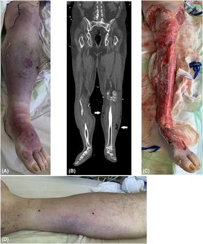
The postoperative status of the patient was precarious with increasing need of hemodynamic support. Noradrenaline infusion up to 0.889 μg/kg/min in addition to fluid administration was required and argipressin at a dosage of 0.02 IE/min was necessary to achieve hemodynamic stabilization. Body temperature was measured 39.1°C and the heart rate at 115 bpm. Laboratory data are described in Table 1. On physical examination, the left limb showed a dusky purplish gangrenous-altered skin with ecchymosis, ruptured violaceous bullae and skin sloughing (Figure 1A). Palpating the edematous skin revealed distinct tenderness and crepitation. Within an hour the skin alterations spread rapidly onto nearly the complete left leg.
| Variable | Reference range, adults | On arrival, ICU | 12 h after arrival, after 1st operation | After 2nd operation, hospital day 3 | Hospital day 5 |
|---|---|---|---|---|---|
| Hemoglobin (mmol/L) | 8.4–10.9 | 5.6 | 5.5 | 5.5 | 5.3 |
| Hematocrit (%) | 36.0–46.0 | 27 | 27 | 25 | 27 |
| White-cell count (per μl) | 3700–9800 | 1620 | 7920 | 6410 | 8600 |
| Platelet count (per μl) | 146,000–328,000 | 83,000 | 45,000 | 44,000 | 35,000 |
| Sodium (mmol/L) | 136–145 | 140 | 140 | 137 | 136 |
| Potassium (mmol/L) | 3.4–4.9 | 3.86 | 4.78 | 5.52 | 4.89 |
| Glucose (mmol/L) | 4.11–5.89 | 8.66 | 9.85 | 12 | 7.42 |
| Creatinine kinase (μmol/L) | <3.2 | 1.05 | 0.73 | 0.31 | 0.24 |
| C-reactive protein (mg/L) | <5 | 109 | 294 | 380 | 273 |
| Procalcitonine (ng/ml) | <0.5 | 56 | >100 | >100 | 38 |
| Creatinine (μmol/L) | 59–104 | 216 | 174 | 173 | 106 |
| Urea nitrogen (mmol/L) | 3.0–9.2 | 37 | 27 | 22 | 12 |
| Lactate (mmol/L) | <2.2 | 3.2 | 3.7 | 3.5 | 2.5 |
| Albumin (g/L) | 35–52 | 27 | 16.8 | 19.4 | 26.1 |
| Aspartate aminotransferase (μmol/L) | 0.17–0.85 | 0.22 | 0.40 | 0.25 | 0.22 |
| International normalized ratio | <1.15 | 68 | 45 | 68 | 91 |
| Activated partial thromboplastin time (s) | <34.4 | 34 | 45 | 68 | 94 |
| pH | 7.37–7.45 | 7.20 | 7.25 | 7.32 | 7.27 |
- Abbreviation: ICU denotes intensive care unit.

Blood cultures were positive with Escherichia coli. A subsequent computed tomography scan depicted gas in the subcutaneous tissue (Figure 1B, arrows). A clinical diagnosis of NF was made and immediate surgical intervention with aggressive epifascial necrosectomy of devitalized tissue and broad-spectrum antibiotic therapy, including meropeneme 1000 mg every 8 h and linezolide 600 mg twice daily, was administered. In addition high-dose (5 mg/mL plasma) intravenous polyspecific immunoglobulin G (IVIG) was dispensed. Histological work-up depicted an extensive acute neutrophilic inflammatory reaction involving the subcutaneous fat and severe necrosis. On the first postoperative day after radical debridement of the left limb (Figure 1C), similar skin alterations developed on the right crus (Figure 1D). A second debridement of the right leg was conducted. The clinical status of our patient deteriorated hereafter rapidly and he succumbed in the course of this severe invasive infection.
3 DISCUSSION
Mortality rates with NF are reported to approximate 20% and remain high despite maximum therapy.3 Severe systemic toxicity occurs more often in patients with older age, immunosuppression, diabetes, malignancy or chronic kidney disease.3 The two most important risk factors associated with increased mortality are delay in surgery and extent of the first debridement.4-6 Other risk factors associated with higher mortality rates are summarized in Table 2.
| Delay in surgery, especially >24 h |
| Extent of first debridement |
Septic condition with hypotension at time of admission
|
Comorbidities
|
Acute kidney injury
|
| Increased serum creatine kinase |
| Decreased international normalized ratio |
| Deceased albumin levels |
| Low hematocrit |
| Older age > 50 years |
| Infection sites apart from extremities |
| Multifocal necrotizing fasciitis |
Common clinical findings include soft tissue edema, tenderness, blisters, bullae formation and a characteristic disproportionate local pain in early stages with transition to pathognomonic skin necrosis with dusky discoloration and crepitus in late stages, which are accompanied by systemic symptoms of shock.7 Wong et al. established a laboratory-based scoring system to help discriminate between necrotizing and non-necrotizing infections, but with its lack of clinical parameters diagnosis cannot be ensured (Table 3).8, 9 Imaging studies contribute to the diagnosis of NF by detecting gas formation in soft tissues. But since there are no specific laboratory markers and radiological proof becomes only available in progressive disease, clinical suspicion is the main clue to the diagnosis of NF.
| Variable | Value | Score | Point value, patient |
|---|---|---|---|
| Hemoglobin (g/dl) |
11–13.5 <11 |
1 2 |
2 |
| White-cell count (per μl) |
15,000–25,000 >25,000 |
1 2 |
0 |
| Sodium (mmol/L) | <135 | 2 | 0 |
| Creatinine (μmol/L) | >141 | 2 | 2 |
| Glucose (mmol/L) | >10 | 1 | 0 |
| C-reactive protein (mg/L) | >150 | 4 | 0 |
| LRINEC score | |||
| ≤5 points: low risk | 4 | ||
| 6–7 points: medium risk | |||
| ≥8 points: high risk | |||
Bilateral NF is an atypical presentation. Concomitant or secondary involvement of remote sites is defined as multifocal NF (MNF) and has been reported in only few cases.10, 11 In a recent study by Lee et al. only 5% of all cases specified as MNF.12 Development of new lesions at distant sites might be caused by either hematogenous dissemination, simultaneous colonization or direct spreading.12, 13 Time lag in manifestation of a subsequent lesion might hint to the underlying mechanism.11 Overall mortality rates of patients presenting with MNF doubles, ranging between 62% and 67%.12
The most likely etiology in this case is the abdominal septic abscess formation, leading to metastatic distribution of E. coli via the blood flow to multiple sites. Our case is particularly rare therein that NF due to E. coli manifests in a multifocal fashion. Such a fulminant presentation of symptoms and progressive disease course is likely because of the compromised immune status of the patient following organ transplantation. This overall situation may also interfere with the laboratory risk indicator for necrotizing fasciitis-score. This fatal outcome emphasizes the urgent necessity of early and aggressive intervention, especially in cases of MNF.
Hallmark for a successful management of NF is immediate extended surgical debridement of all affected tissues. Concomitantly initiation of broad-spectrum antibiotic coverage e.g. with piperacillin/tazobactam to also address anaerobic bacteria plus clindamycin as soon as NF is suspected is essential.
Adjuvant therapeutic approaches may include administration of intravenous immunoglobulins, especially in the early disease course and in the setting of streptococcal infection belonging to group A.14-16 In the index case IVIG was administered because of proven immunoglobulin deficiency (IgG 3.07 [7–16] g/L) in the immunocompromised patient. Hyperbaric oxygen therapy (HBOT) as another adjunctive therapeutic option is discussed controversially.17, 18 We did not implemented HBOT in this case, since we lacked the possibility for a pressure chamber with affiliated intensive care capacity. Moreover transportation risks for our patient were assessed to be too high at the time.
4 CONCLUSION
Necrotizing fasciitis diagnosis is often delayed because of the lack of specific clinical features in the initial stage of the disease. Clinicians should be aware of NF as differential diagnosis especially if there are indications of soft tissue infection combined with signs of systemic involvement. NF might rarely present in a multifocal manner, which tends to have an even more unfavorable outcome.
AUTHOR CONTRIBUTIONS
Sascha T. Bender: Conceptualization; resources; writing – original draft; writing – review and editing. Peter R. Mertens: Supervision; writing – review and editing. Christian Gross: Data curation; supervision; writing – original draft.
ACKNOWLEDGMENTS
None. Open Access funding enabled and organized by Projekt DEAL.
CONFLICT OF INTEREST
The authors declare that they have no competing interests.
CONSENT
Written consent has been obtained from the patient.
Open Research
DATA AVAILABILITY STATEMENT
Data sharing not applicable.


