Volume 11, Issue 1 e6869
CASE IMAGE
Open Access

Radiological manifestation of optic nerve infarction in a patient with invasive mucormycosis secondary to diabetic ketoacidosis in Pakistan

Shehroze Tabassum

Shehroze Tabassum

Department of Internal Medicine, King Edward Medical University, Lahore, Pakistan

Contribution: Conceptualization, Writing - original draft, Writing - review & editing

Search for more papers by this author
Aroma Naeem

Aroma Naeem

Department of Internal Medicine, King Edward Medical University, Lahore, Pakistan

Contribution: Writing - original draft

Search for more papers by this author
Afshan Shakir

Afshan Shakir

Department of Radiology, King Edward Medical University, Lahore, Pakistan

Contribution: Writing - original draft, Writing - review & editing

Search for more papers by this author
Faiza Afzal

Faiza Afzal

Department of Radiology, King Edward Medical University, Lahore, Pakistan

Contribution: Writing - original draft

Search for more papers by this author
Laya Ohadi

Corresponding Author

Laya Ohadi

School of Medicine, Shahid Beheshti University of Medical Sciences, Tehran, Iran

Correspondence

Laya Ohadi, School of Medicine, Shahid Beheshti University of Medical Sciences, Tehran, Iran.

Email: [email protected]

Contribution: Writing - original draft

Search for more papers by this author
First published: 19 January 2023

Abstract

A 35 years old male patient presented in the hospital with complaints of left-sided facial swelling, blindness in the left eye, and left eye proptosis. He had a concomitant history of diabetic ketoacidosis. Magnetic resonance imaging was advised, which revealed infected tissue of the left cheek, optic nerve infarction, intracranial extension, and leptomeningeal involvement by the disease process.

1 CLINICAL IMAGE AND DESCRIPTION

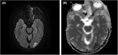
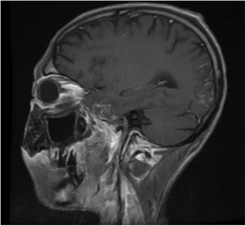
A 35 years old male patient presented in the hospital with complaints of left-sided facial swelling, blindness in the left eye, and left eye proptosis. He had a concomitant history of diabetic ketoacidosis (DKA). Magnetic resonance imaging (MRI) was advised. MRI findings were suggestive of rhino occulo cerebral mucormycosis. Mucor, a fungus, can lead to deadly infection of the paranasal sinuses, eventually involving the orbit and brain.1 There were only limited cases of diffusion-weighted MRI demonstrating ischemic optic neuropathy when the literature was scoured.2 Figures 1-3 with description provide a comprehensive illustration.
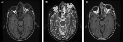
Details are in the caption following the image
(A) Time to echo phase 1 (T1WI) shows left optic nerve thickening with no altered signals. (B) T2WI shows left optic nerve with altered T2 signals and shows mild thickening. (C) T1W Post-contrast shows slight perineural enhancement with optic nerve thickening.
Details are in the caption following the image
(A, B) Diffusion-weighted imaging (DWI). (A) shows hyperintense signals within the left optic nerve, which correlates with hypointense signals on apparent diffusion coefficient (ADC) mapping (B), suggesting diffusion restriction consistent with optic nerve infarction. Further diffusion restriction is seen in the left occipital lobe suggesting an intracranial extension of the disease process.
Details are in the caption following the image
Time to echo phase 1 (T1WI) with contrast reveals interrupted leptomeningeal enhancement in left temporal and occipital lobes suggesting meningitis – sequelae of mucormycosis suggesting leptomeningeal involvement by the fungal infection.

AUTHOR CONTRIBUTIONS

Shehroze Tabassum: Conceptualization; writing – original draft; writing – review and editing. Aroma Naeem: Writing – original draft. Afshan Shakir: Writing – original draft; writing – review and editing. Faiza Afzal: Writing – original draft. Laya Ohadi: Writing – original draft.

ACKNOWLEDGEMENT

None.

    CONFLICT OF INTEREST

    None.

    CONSENT

    Written informed consent was obtained from the patient for publication of this image report in accordance with journal's patient consent policy.

    DATA AVAILABILITY STATEMENT

    The data and materials used in the current study are available from the corresponding author upon reasonable request.

      The full text of this article hosted at iucr.org is unavailable due to technical difficulties.