Intraosseous lipoma of Calcaneum: A rare incidental finding
Abstract
Intraosseous lipoma is a rare benign lesion of bone. We present a case of an intraosseous lipoma of the calcaneum detected as an incidental finding, as a well-delineated osteolytic lesion with the central area of calcification, on plain radiography. Diagnosis can be done with a computed tomography scan and/or magnetic resonance imaging.
1 INTRODUCTION
Lipomas are benign lesions of adipose tissue. Intraosseous lipomas are rare entities, accounting for less than 0.1% cases of all primary bone tumors.1 They can occur anywhere in the skeleton; the common location being the lower limbs including the calcaneum. Some cases of intraosseous lipomas are asymptomatic which are detected incidentally on radiographs performed for evaluation of some other disorders while others may have symptoms like heel pain. These lesions are frequently misdiagnosed for other conditions. Diagnosis with a plain radiograph alone may be difficult, and the use of computed tomography (CT) scan and/or magnetic resonance imaging (MRI) can easily confirm the condition. We present a case of an incidental intraosseous lipoma diagnosed by a CT scan in a 36-year-old male.
2 CASE PRESENTATION
A 36-year-old male patient, a military person, presented with mild pain and swelling over the left ankle region for 2 days following the history of mechanical trauma sustained while running. Clinical examination revealed mild tenderness in the lateral aspect of the ankle, without evidence of a palpable mass. Ankle movements were normal except for mild pain on inversion. He was a social drinker and non-smoker. He had no history of chronic medical conditions and was also not under any long-term medications. He was initially managed with oral analgesics and advised to refrain from involving in strenuous exercises like running.
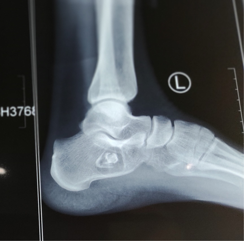
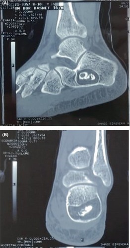
A plain radiograph of the left ankle was ordered which showed a well-defined, radiolucent bone lesion of size measuring about 24 × 28 mm within the anterior part of the calcaneum, with central radiopaque area of calcification called as Cockade sign as shown in Figure 1. Subsequently, a CT scan of the left ankle was performed to characterize the lesion, revealing a well-defined lytic lesion in the calcaneal neck measuring 25 × 29 × 34 mm with a smooth sclerotic margin and narrow transition zone. It showed areas of low attenuation equivalent to fat density (−60 Hounsfield units) and coarse calcification in the center. The inferior cortex of the calcaneum was thinned out without any cortical break as shown in Figure 2A,B. A diagnosis of intraosseous lipoma (stage 3) of the left calcaneum was thus made.


3 DISCUSSION
Intraosseous lipomas are very rare benign tumors of bones with an incidence of less than 0.1% of all primary bone tumors mostly affecting the bones of lower extremities with calcaneum being a common location like in our case.2 Other long bones femur, tibia, and fibula are also affected in metaphyseal and diaphyseal regions. They are mostly solitary and unifocal. Lipomas are derived from mature adipocytes, but the exact etiology is poorly understood. Many believe in a primary origin from bone marrow fat, while others support trauma and fat degeneration, infective pathologies, and fat metaplasia. Age and sex predilection are also not fully agreed upon. Several cases are asymptomatic and detected as incidental findings while working on some other conditions. Symptomatic cases may present with pain, swelling, and tenderness.3 Intraosseous lipomas can undergo changes like fat necrosis, cystic degeneration, dystrophic calcification, fibrosis, and bone infarction. Malignant transformation has been documented in very few cases of intraosseous lipomas.4
Diagnosis with simple plain X-ray alone is difficult for an intraosseous lipoma; the presence of benign-appearing osteolytic bone lesion with well-defined margins with intralesional calcification commonly referred to as Cockade sign may be a useful clue.5 With the advent of CT and MRI, we can detect the nature of the lesion without the need for biopsy or surgical excision.6 However, it is important to understand histopathology to better recognize the imaging features of intraosseous lipomas. Based on the histopathological features, intraosseous lipomas are divided into three types: Stage 1 includes solid tumors of viable lipocytes; stage 2 includes transitional cases with partial fat necrosis and focal calcification but with areas of viable lipocytes; and stage 3 includes advanced cases where fat cells have died with varying degree of changes like calcification, cyst formation, and reactive new bone formation.7 The radiological features including CT scan findings correspond to the histologic stages of the lesion. Stage 1 lesions are lucent representing viable, non-necrotic fat with resorption of bony trabeculae; stage 2 lesions have lucent areas of viable fat along with radiodense areas of fat necrosis and dystrophic calcification and may be expansile too. Stage 3 lesions are heterogenous, more radiodense due to calcification and extensive fat necrosis along with the thick sclerotic border, and may show cystic changes within.8
MRI is also an excellent method for the identification of intraosseous lipomas by visualizing fat within the lesions.2 Stage 1 lipomas show viable fat isointense to subcutaneous fat on T1-weighted sequences and show low signal intensity with fat suppression on T2-weighted images. A thin surrounding circumferential rim consistent with reactive sclerosis with low signal intensity on T1- and T2- weighted sequences demarcating the margin of the fatty lesion may be present. Stage 2 lesions also show fat and the circumferential rim of decreased signal on T1- and T2-weighted images along with the low-signal-intensity areas in the central portion of the lesion consistent with calcifications. Stage 3 lesions have a thin peripheral rim of fat, central calcification, and a thick rim of surrounding sclerosis with low signal intensity on T1- and T2-weighted sequences. A variable signal is seen on T1-weighted and an increased signal is seen on T2- weighted images in areas of fat necrosis.9
The differential diagnoses for this condition include bone infarct, unicameral bone cyst, aneurysmal bone cyst, chondromyxoid fibroma, fibrous dysplasia, osteoblastoma, giant cell tumor, and liposclerosing myxofibrous tumors (LSMFT).9 Clinicoradiological correlation is essential to determine the diagnosis of intraosseous lipoma. Most cases can be managed conservatively. The prognosis is very good, and the chance of recurrence is negligible. The possible surgical indications for management can be the presence of a painful lesion, pathological fracture, necessity for histological diagnosis or to decrease the risk of malignant transformation.10, 11 Common surgical options are curettage with or without bone grafting, or with a bone graft substitute.
4 CONCLUSION
Calcaneal intraosseous lipomas are rare benign conditions of bone that may be detected incidentally and may pose a diagnostic challenge. Evaluation using CT scan and/or MRI can be used for the definitive diagnosis in addition to the plain radiographic images without the need for biopsy and histopathological examination. These lesions can generally be managed conservatively and have an excellent prognosis.
AUTHOR CONTRIBUTIONS
Bishesh Lamichhane: Conceptualization; data curation. Saral Lamichhane: Conceptualization; writing – original draft. Roshan Shah: Data curation; writing – review and editing. Mamata Yadav: Data curation; writing – review and editing. Sujata Pant: Project administration; supervision; writing – review and editing.
ACKNOWLEDGEMENTS
None.
FUNDING INFORMATION
None.
CONFLICT OF INTEREST
There are no conflicts of interest to declare.
ETHICAL APPROVAL
None.
CONSENT
Written informed consent was obtained from the patient to publish this report in accordance with the journal's patient consent policy.
Open Research
DATA AVAILABILITY STATEMENT
Data available on request from the authors.


