Imaging manifestations of small cell neuroendocrine carcinoma of the ureter
Abstract
Small cell neuroendocrine carcinoma (SCNEC) of the ureter is a rare malignant tumor originating from the metaplasia of urothelial cells. This report presents a case of ureteral SCNEC that was preliminarily disclosed by computed tomography; thereafter, transabdominal ultrasonography, transrectal ultrasonography, and magnetic resonance urography were performed to characterize the mass.
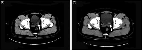
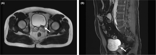
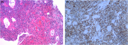
A 35-year-old man with a history of backache and hematuria over 1 week was admitted to our hospital. Transabdominal ultrasonography revealed dilation of the left renal collecting system and upper ureter due to blockage of the lower ureter by a hypoechoic mass. Coincidentally, the mass was located in the pelvic cavity next to the rectum. Transrectal ultrasonography was performed; a thickened lower ureter wall, whose structure was disordered and stiff, and additional details of the mass were obtained (Figure 1). Two days ago, abdominal computed tomography (CT) explored a soft tissue mass of the lower ureter that should be distinguished from tumors in other hospitals (Figure 2). Furthermore, magnetic resonance urography (MRU) suggested that the ureteral mass had a low signal filling defect, which was considered a carcinoma due to the lymph nodes being scattered around it (Figure 3). Ultimately, the patient underwent cystoscopy and surgical excision. Finally, pathological examination performed confirmed small cell neuroendocrine carcinoma (SCNEC) (Figure 4).


Small cell neuroendocrine carcinoma of the ureter is a highly malignant tumor originating from the metaplasia of urothelial cells with or without neuroendocrine function.1 Due to its rare nature, ureteral SCNEC is always misdiagnosed or escapes diagnosis in the early stage and has a poor prognosis in the later stage on imaging.2 However, SCNEC is usually detected by CT but rarely by ultrasonography and MRU, which could provide more features of ureteral SCNEC, including its composition, pattern, and blood flow. Imaging manifestations of inverted urothelial papilloma is a noninvasive endophytic urothelial neoplasm occurred in the renal pelvis, ureter, and urinary bladder, which shows iso-intense on T1-weighted images and either iso-intense or slightly higher in intensity on T2-weighted images.3 However, it is not uncommon to mistake SCNEC as other ureteral urothelial carcinomas depending on ultrasonography and MRU, especially high-grade ureteral urothelial carcinoma. In conclusion, abdominal CT cannot be avoided and cystoscopy remains the diagnostic procedure of choice.4
AUTHOR CONTRIBUTIONS
Guiwu Chen wrote the original draft of this clinical image and made the subsequent revisions. Xiong Huang participated in the computed tomography analysis and interpretation. Wenqin Liu and Zhizhong He participated in the ultrasonography and magnetic resonance urography analysis and interpretation. Xiaomin Liao participated in the pathology image analysis and interpretation. Yuhuan Xie assisted in the revision and supervised the overall production of this report.
ACKNOWLEDGMENT
None.
FUNDING INFORMATION
No funding was received for this study.
CONFLICTS OF INTEREST
The authors declare that there is no conflict of interest.
CONSENT
Written informed consent was obtained from the patient to publish this report in accordance with the journal's patient consent policy.
Open Research
DATA AVAILABILITY STATEMENT
The data used to support the findings of this study are available from the corresponding author upon request.


