Primary pleural lymphoma accompanied by silicosis
Abstract
Reports of malignant lymphoma accompanied by silicosis are limited. A 93-year-old man with silicosis presented with right massive pleural effusions and was diagnosed with primary pleural lymphoma. Since there was no evidence of chronic pyothorax or Epstein–Barr virus infection, it may be due to silicosis-associated chronic inflammation.
1 INTRODUCTION
Although lung cancer is the most common malignancy associated with silicosis, there are few reports of malignant lymphoma accompanied by silicosis.1 To the best of our knowledge, there is only one case report of primary pleural lymphoma associated with silicosis.2 We report a case of pleural lymphoma in a patient with silicosis presented with a massive pleural effusion.
2 CASE PRESENTATION
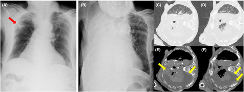
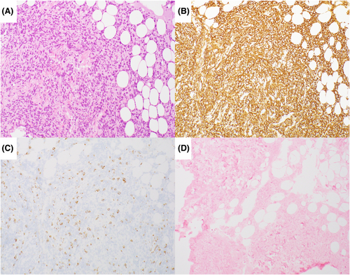
A 93-year-old man with a history of silicosis was referred for progressive dyspnea. Chest imaging revealed right massive pleural effusions and multiple pleural tumors, in addition to small silicotic nodules and enlarged hilar and mediastinal lymph nodes with calcification (Figure 1). Plasma levels of soluble interleukin-2 receptors were elevated (2770 U/ml), and malignant cells were observed in the pleural fluid. Thoracoscopy performed under local anesthesia revealed multiple protruding papillary lesions in the pleura, and diffuse large B-cell lymphoma was confirmed pathologically (Figures 2 and 3). Brain and abdominal to pelvic computed tomography showed no tumor lesions, and the patient was diagnosed with primary pleural lymphoma. The patient died 16 days after palliative treatment owing to poor general condition.


3 DISCUSSION AND CONCLUSION
Complications associated with silicosis include lung cancer and tuberculosis; however, the association of silicosis with malignant lymphoma is unclear. Primary pleural lymphoma is known to be associated with chronic tuberculous pyothorax, Epstein–Barr virus (EBV) infection, and autoimmune diseases.3 In this case, chronic pyothorax was not observed, and EBV was not detected on pathological examination; therefore, we considered the disease onset may have been due to chronic inflammation by silicosis. Since silicosis is rarely accompanied by pleural effusions,4 when pleural effusions are found in patients with silicosis, lymphoma should be considered, in addition to lung cancer and tuberculosis.
AUTHOR CONTRIBUTIONS
Y.M. wrote the initial draft of the manuscript. A.O. and K.I. were responsible for the drafting and image modification. A.O. performed the thoracoscopy. All authors read and approved the final manuscript.
ACKNOWLEDGMENT
None.
FUNDING INFORMATION
This research was not supported by any specific grant from any funding agency in the public, commercial, or non-profit sectors. Therefore, no funding body was not involved in the design of the study, the collection, analysis, and interpretation of the data, the writing of the manuscript, or the decision to submit the manuscript for publication.
CONFLICT OF INTEREST
None.
CONSENT
The patient has passed away. Thus, written informed consent to publish this report was obtained from the patient's relatives before the submission process.
Open Research
DATA AVAILABILITY STATEMENT
No datasets were generated or analyzed during this case report.


