A rare case of a laryngeal angiomyolipoma
Level of evidence: 4.
Patient seen and treated at Park West Hospital by Nicholas Panella, M.D.
Abstract
Laryngeal angiomyolipoma is a rare tumor with few reported cases in the literature. The case report explains a 62-year-old man who presents with dyspnea and found to have a laryngeal angiomyolipoma staining CD34 positive, but HMB45 and Melan-A negative.
1 INTRODUCTION
Angiomyolipoma is a rare benign tumor often found in the kidney in conjunction with tuberous sclerosis. An angiomyolipoma of the larynx is rare with only five reported cases in the literature. Diagnosis is typically made with immunohistochemistry, with an angiomyolipoma staining positive for a smooth muscle marker such as desmin or SMA and a melanocytic marker such as HMB45. This tumor is important to be on the differential for common but chronic ENT complaints of dyspnea and hoarseness as well as a possible complication of intubation.
2 CASE REPORT
A 62-year-old man presented as an ICU transfer from a small community hospital with complaints of 3 days of worsening dyspnea and O2 saturation of 84%. He had occasional shortness of breath and episodic throat clearing for 9 months prior. His medical history was unremarkable.
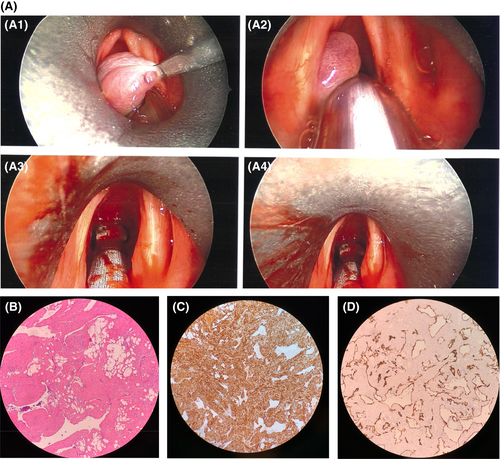
Flexible laryngoscopy was performed, and a large pedunculated glottic mass was visualized coming from the immediate subglottis with ball valving movement with potential for complete airway obstruction. The patient was taken emergently to surgery where the tumor was excised via direct laryngoscopy with microlaryngeal dissection with only minor difficulty intubating (Figure 1). On final pathology, the diagnosis of laryngeal angiomyolipoma was made based on histologic appearance (Figure 1).

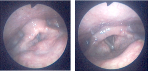
The patient was discharged on omeprazole daily to assist healing. At 3-week follow-up in the clinic, his voice had returned to normal and he was having no persistent dyspnea. Flexible laryngoscopy was performed and vocal folds were examined with no edema and no persistent mass (Figure 2).

3 DISCUSSION
Angiomyolipoma is a rare benign tumor composed of adipose tissue, smooth muscle, and blood vessels most commonly found in the kidney as a result of sporadic or inherited mutation of TSC1 or TSC2. Sporadically, this occurs more in middle-aged adults, while the inherited type is due to tuberous sclerosis seen in the younger population. Tuberous sclerosis causes a multitude of benign tumors that can be accompanied by other symptoms, most commonly epilepsy and developmental delay.
With only five known cases of laryngeal angiomyolipoma, the location of this tumor is rare. When comparing with other laryngeal angiomyolipoma cases in the literature, five of six are found in adult men.1-4 Contrasted with sporadic angiomyolipoma in all locations, female predominance is cited at 4:1.5 It is not known why these tumors have been more commonly reported in the male larynx, but further investigation is warranted as future cases are reported.
Pathological inspection and immunohistochemical staining are helpful to make the diagnosis of angiomyolipoma. The mass discussed was myosin and CD34 positive and HMB45/Melan-A negative. All but one of the laryngeal angiomyolipoma cases in the literature are smooth muscle marker positive (SMA, desmin), and half are positive for the melanocytic marker HMB45.1-4 In one case of angiomyolipoma of the tongue, the mass was found to be both SMA and HMB45 positive.5 Several cases of angiomyolipoma outside the kidney are negative for HMB45.6 This difference is possibly due to the difference of spindle cells versus epithelioid cells within the tumor. The final diagnosis was ultimately based upon histologic examination, because negative HMB45 staining typically indicates a diagnosis other than AML.3
The patient had been transferred from a smaller hospital with less experience in laryngeal cancers and it was assumed that the mass was a laryngeal squamous cell carcinoma based on CT findings.7 Upon bedside visualization of the mass, granuloma, chondroma, PeCOMA, and angioleioma were differential diagnoses. These are all masses that are more commonly seen in the larynx and can cause symptoms of dyspnea and airway obstruction. Malignancy did not seem as likely because of the well-circumscribed nature of the mass and lack of ulceration. In order to make the diagnosis of angiomyolipoma, pathologic inspection was required. Additionally, size of the mass should guide excision, with most being excised by direct laryngoscopy with endolaryngeal microsurgery.
This is an important finding not only for otolaryngologists, but also for all providers because of the possibility of airway compromise. In one reported case, the laryngeal angiomyolipoma was found incidentally during an upper gastrointestinal endoscopy.3 Such a lesion can present a challenge during intubation and is therefore relevant to anesthesiologists and intensivists. In earlier stages of tumor growth, patients often present with common symptoms of dyspnea, stridor, voice change, and swallowing discomfort. Because of this, it is important to keep this rare tumor on the differential even with routine ENT complaints.
4 CONCLUSION
Laryngeal angiomyolipoma is a rare tumor that should not be overlooked as a possible diagnosis. Immunohistochemical staining of smooth muscle and melanocytic markers could be helpful for confirmation but is not necessarily consistent throughout the five previously published cases of laryngeal angiomyolipomas.
SUMMARY FOR LAY READERSHIP
A case report of a patient presenting to the emergency department with complaints of dyspnea and is found to have an angiomyolipoma of the larynx.
AUTHOR CONTRIBUTIONS
Lillian McCampbell: Conceptualization; writing – original draft. Mark Williams: Validation; writing – review and editing. Nicholas Panella: Resources; supervision; writing – review and editing.
ACKNOWLEDGMENT
None.
FUNDING INFORMATION
No source of financial support or funding.
CONFLICT OF INTEREST
All authors have no conflict of interest.
CONSENT
Written patient consent has been signed and collected in accordance with the journal's patient consent policy.
Open Research
DATA AVAILABILITY STATEMENT
Data sharing not applicable to this article as no datasets were generated or analysed during the current study.


