Fatal invasive gastric mucormycosis: Two case reports
Abstract
Mucormycosis is a fungal infection affecting most commonly immunocompromised patients. Hereby, we report two cases: the first one is about a 61-year-old female with diabetes who presented with vomiting. The upper gastrointestinal endoscopy showed a budding grayish process which corresponded to an invasive mucormycosis in histology. As laboratory tests showed renal dysfunction, conventional amphotericin B was started at low doses since liposomal form was unavailable in Tunisia. Evolution was marked by a worsening of renal function leading to drug therapy withdrawal. Total gastrectomy was delayed because of a pulmonary embolism and was practiced 2 months later. The patient passed away 10 days after surgery. The second patient was a 59-year-old man who presented with vomiting and fast worsening of general state. At admission, he had a septic shock. Explorations revealed an invasive gastric mucormycosis. He died few days after admission. Thus, prompt diagnosis of mucormycosis and rapid initiation of treatment based on amphotericin B and surgical debridement is necessary to improve prognosis.
1 INTRODUCTION
Mucormycosis or black fungus is an uncommon fungal infection that affects usually immunocompromised patients such as patients with diabetes, renal dysfunction, cancer, or under immunosuppressive therapy. It is due to a non-septate filamentous fungus that affects often facial structures (nose, sinuses, eye, and brain), lungs, or skin and less commonly the gastrointestinal tract, the stomach being on the top of the list. The major route of infection transmission is inhalation. However, when it concerns the gastrointestinal tract, ingestion of filaments is incriminated. The clinical signs usually depend on the site of origin. Treatment is based on amphotericin B and in most cases surgical debridement of necrotic tissues, though mucormycosis is associated with poor prognosis and high rates of mortality.1
2 CASES PRESENTATION
2.1 Case one
A 61-year-old Tunisian woman with type 2 diabetes mellitus, hypertension, dyslipidemia, and family history of peptic ulcer presented to our gastroenterology department with a 7-day history of vomiting preceded by a 4-month history of dry cough for which she took inhaled corticosteroids. On physical examination, she had no fever, her body mass index was 29.8 kg/m2. Her blood pressure was 120/70 mmHg. Her abdominal exam was normal. Initial laboratory tests showed renal dysfunction with creatinine level at 163 μmol/L and creatinine clearance at 60 ml/min, hemoglobin was 11.5 g/dl, white blood cell count (WBC) 11,100 cells/μl, C-reactive protein (CRP) 132 g/L, and blood sugar 20 mmol/L (Table 1).
| Laboratory test | Results | Reference range |
|---|---|---|
| Urea (mmol/L) | 15.5 | 2.5–7.6 |
| Creatinine (μmol/L) | 163 | 50–100 |
| Clearance (ml/min) | 30 | >90 |
| Hemoglobin (g/dL) | 11.5 | 12–17 |
| WBC (cells/mm3) | 11,100 | 4500–10,000 |
| Platelets (cells/mm3) | 252,000 | 150,000–400,000 |
| CRP (g/L) | 132 | <6 |
| Sodium (mmol/L) | 134 | 136–145 |
| Albumin (g/L) | 29 | 38–46 |
| Blood sugar (mmol/L) | 20 | 3.5 - 6.1 |
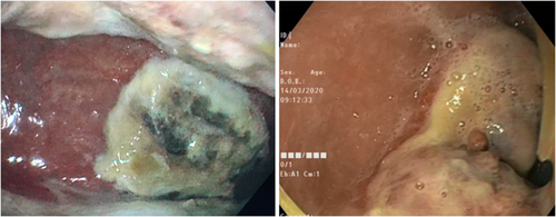
Upper gastrointestinal endoscopy revealed budding and infiltrating grayish process resting on an ulcerated fundic mucosa (Figure 1).
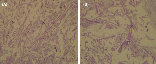
Anatomopathological exam concluded invasive mucormycosis by demonstrating zygomycosis hyphae without any malignant signs (Figure 2).
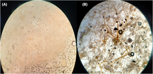
These findings were confirmed by parasitological examination (Figure 3).
A brain-face-chest-abdomen and pelvis computed tomography and ENT exam were practiced to search other sites of mucormycosis (Figure 4). They confirmed the presence of other infection site.
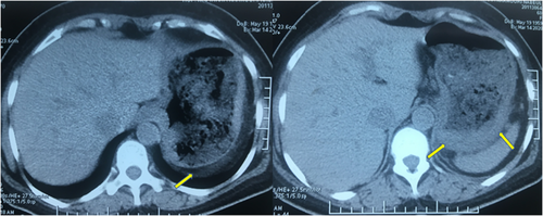
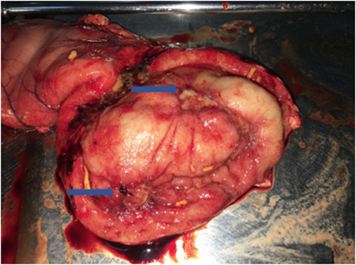
Since the liposomal form of amphotericin B which is less nephrotoxic was not available in our country, therapy based on conventional amphotericin B was started on the fifth day of diagnosis at progressive dose 5 mg/day on day one until 25 mg/day (0.4 mg/kg/day) on day 5. Evolution was marked by an aggravation of renal dysfunction: creatinine level increased to 208 μmol/L and creatinine clearance decreased to 25 ml/min. Thus, we stopped antifungal therapy. In day 19, she was programmed for a total gastrectomy but presented a respiratory distress due to pulmonary embolism. The patient underwent total gastrectomy 2 months after diagnosis. The examination of the stomach showed two digging ulcers in the fundus (Figure 5). The patient died 10 days after surgery.
2.2 Case two
A 59-year-old man originally from Cango living in Tunisia without any medical history presented to the hospital with gastric pain and vomiting and a fast worsening of the general state. At admission, he had fever. He had low blood pressure (80/45 mmHg) and tachycardia (110 BPM). Abdominal exam showed epigastric tenderness and a moderate ascites. Laboratory findings revealed severe normocytic anemia with 7.1 g/dl hemoglobin, elevated WBC count (24,900 cells/μl [93% neutrophils]), and thrombopenia at 72,000/mm3. He had high CRP (231 g/L) with positive procalcitonin test (2.53 ng/ml). We concluded a septic shock, and the patient had broad-spectrum antibiotherapy and vascular filling with vasoactive drugs.
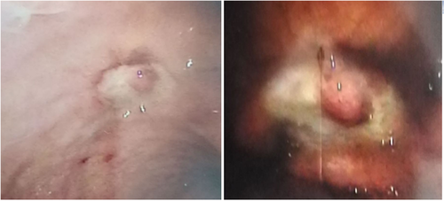
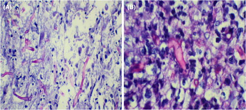
Upper gastrointestinal endoscopy revealed a very fragile mucosa and a gastric ulcer with bleeding stigmata (Figure 6). Abdominal ultrasound was normal besides moderate ascites. Anatomopathological exam showed typical broad zygomycetes hyphae (Figure 7).
Six days later, the patient developed an hemophagocytic syndrome confirmed by cytological examination of the bone marrow. Evolution was rapidly fatal with continuous decrease in hemoglobin level despite transfusions, and the patient died within 8 days after admission.
3 DISCUSSION
Zygomycosis was first described as a cause of human disease in 1885.2 Unlike other fungal opportunist infections affecting patients with compromised immunity, cancer, or recipients of organ transplant, mucormycosis can occur in patients with diabetes mellitus,2-4 renal insufficiency, alcoholism,5-7 or even with an intact immunity system.8-10 These underlying conditions not only predispose to the disease but also worsen the outcome of patients. Our first patient had diabetes and renal insufficiency as risk factors of mucormycosis.10
The mode of transmission of mucormycosis involves inhalation or ingestion of spores and direct inoculation into damaged mucocutaneous surfaces.9
The common sites of infection are sinuses(39%) which constitute the predilected infection site in diabetic patients, skin (19%) in particular in patients with no risk factor, and lungs (24%) in patients with cancer.2, 11
The gastrointestinal tract is less commonly affected (7%) with 67% of stomach infection and 25% of intestine infection.2, 10 Factors predisposing for gastrointestinal mucormycosis (GIM) include malnutrition, typhoid fever, uremia, and penetrating trauma.9, 12
The clinical manifestations of GIM include fever, abdominal pain, nausea, vomiting, diarrhea, hemorrhage, or gastrointestinal perforation.9, 13 Our first patient presented only vomiting, and the second patient presented with fever, abdominal pain, and vomiting. The confirmation of diagnosis is based on anatomopathological examination of tissue samples showing fungal hyphae.9, 14
GIM is usually fatal with 85% of mortality.2, 11, 15 The mainstay of therapy in GIM is a combination of surgical debridement and intravenous antifungal therapy. Early initiation of antifungal therapy improves survival and can even exempt the patient from surgery in case of non-invasive form of the disease.11 A delay in treatment greater than 6 days worsens the prognosis and seems to double the mortality rates.9 The cornerstone of drug therapy is amphotericin B which constitutes the first line treatment at a daily dose of 1 to 1.5 mg/kg for a duration of 4–6 weeks.5, 9, 16 The liposomal form is preferred for its lower risk of nephrotoxicity and the possibility to administrate higher doses: 5 to 10 mg/kg/day.2, 17, 18 Posaconazole and isavuconazole are also effective agents and can be used as second-line therapy. Surgical debridement of necrotic tissues is often required for invasive mucormycosis.1, 18 In our first patient, liposomal form of amphotericin B was not available in our country; thus, we started treatment with conventional amphotericin B at low doses which was stopped at the 5th day because of worsening of renal dysfunction, and we opted for the surgical debridement. As for the second patient, he did not receive this treatment since he was in a septic shock.
4 CONCLUSION
Gastric mucormycosis is a rare entity that affects usually predisposed patients such as immunocompromised, alcoholic, and diabetic patients. However, immunocompetent patients are not spared. Its treatment is based on amphotericin B and surgical debridement of all necrotic tissues. The mortality rates remain high up to 85%. Thus, early diagnosis and prompt therapy are necessary to improve prognosis.
AUTHOR CONTRIBUTIONS
Have made substantial contributions to conception and design, or acquisition of data, or analysis and interpretation of data: Manel Moalla, Salwa Nechi, Amina Bani, Aicha Elloumi, Sana Jemal. Been involved in drafting the manuscript or revising it critically for important intellectual content: Amal Khsiba, Manel Moalla. Given final approval of the version to be published. Each author should have participated sufficiently in the work to take public responsibility for appropriate portions of the content: Mohamed Moussaddek Azouz. Agreed to be accountable for all aspects of the work in ensuring that questions related to the accuracy or integrity of any part of the work are appropriately investigated and resolved: Amal Khsiba, Manel Moalla, Mohamed Moussaddek Azouz.
ACKNOWLEDGMENTS
None.
FUNDING INFORMATION
None.
CONFLICT OF INTEREST
We have no conflict of interest to declare.
ETHICAL APPROVAL
Patient personal data have been respected.
CONSENT
Written informed consent was obtained from the patients to publish this report in accordance with the journal's patient consent policy.
Open Research
DATA AVAILABILITY STATEMENT
Data openly available in a public repository that issues datasets with DOIs.


