Hiatal hernia presenting as a left atrial mass
Abstract
Cardiac masses pose diagnostic challenges. We present a 62-year-old woman presented for evaluation of chest pain and palpitations. Transthoracic echocardiography showed left atrial mass, which was subsequently identified as hiatal hernia by cardiac magnetic resonance imaging.
1 CASE PRESENTATION
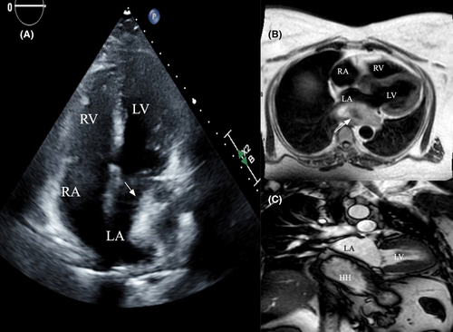
A 62-year-old Caucasian woman with a history of hypertension presented for evaluation of palpitations and chest pain. She had a transthoracic echocardiogram which showed an ejection fraction of 60%–65% and a large hyperechoic mass within the left atrium near the posterior wall (Figure 1A). Labs were within normal limits. She underwent cardiac magnetic resonance imaging (MRI) which revealed large hiatal hernia along the inferior and lateral aspect of the left atrium resulting in compression of the left atrium. It measures around 8.1 cm × 4.3 cm in the sagittal views (Figure 1B,C). No other masses were noted in the left atrium/other heart chambers. She underwent surgical correction and her symptoms resolved completely.

2 DISCUSSION
Cardiac masses pose diagnostic challenges. Not all left atrial masses are cardiac in nature. Echocardiography is the initial diagnostic tool and computed tomography (CT) and MRI will further aid in the diagnosis of the cardiac masses.1 Hiatal hernia can present as cardiac mass depending on the size of the hernia protruding into the chest and can present with cardiac symptoms such as palpitations due to atrial arrhythmias and chest pain due to gastro esophageal reflux.2 Multimodality imaging helps to identify the cardiac masses accurately.
AUTHOR CONTRIBUTIONS
JK collected the patient data, literature review, and wrote the manuscript; JP collected the patient data and revised the manuscript. All authors have read and approved the final version of the manuscript.
ACKNOWLEDGEMENT
None.
CONFLICT OF INTEREST
The authors declare no conflicts of interest.
CONSENT
Written informed consent was obtained from the patient to publish this report in accordance with the journal's patient consent policy.
Open Research
DATA AVAILABILITY STATEMENT
All data generated or analyzed during this study are included in this manuscript.


