Recurrent cervical osteomyelitis after radiation therapy in a patient with oropharyngeal cancer
Funding information
None.
Abstract
It is crucial to consider cervical osteomyelitis as a differential diagnosis for neck pain in patients who underwent radiotherapy for early diagnosis and management, thereby preventing the development of potentially debilitating neurologic symptoms.
1 CASE DESCRIPTION
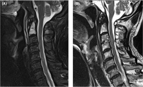
A 67-year-old man with a 10-year history of oropharyngeal cancer treated with chemoradiotherapy (70 Gy) presented with a 5-week history of fever and neck pain. Past medical history revealed cervical osteomyelitis (C1/C2) 4 years ago (Figure 1A). He was afebrile and had normal vital signs. Physical examination showed limitation of cervical range of motion in all directions. Laboratory examination demonstrated an elevated erythrocyte sedimentation rate (30 mm/h) without leukocytosis. Cervical magnetic resonance imaging revealed a new deformity at the C3-C4 vertebral endplates with hyperintense signals at C3/C4 intervertebral disks on T2 (Figure 1B). He was diagnosed with another episode of cervical osteomyelitis. Empirical treatment with vancomycin was started after collecting cultures.

Although only 14% of all vertebral osteomyelitis cases involve the cervical spine, cervical osteomyelitis has the highest risk for neurologic complications (ie, motor weakness or paralysis).1 High-dose radiation therapy for primary head and neck malignancies is a known risk factor for osteomyelitis at the irradiated site.2 The pathophysiologic mechanisms of radiation-induced osteomyelitis include osteoblast and osteoclast inhibition, vascular and lymphoid tissue damage, and mucosal ulceration, resulting in increased susceptibility to infection.2
Neck pain in patients with previous radiation therapy should be evaluated for osteomyelitis to prevent debilitating neurologic symptoms.
AUTHOR CONTRIBUTION
YH examined and treated the patient. TT read the MRI. Both authors wrote the manuscript and approved it for publication.
ACKNOWLEDGEMENTS
Nothing to disclose.
CONFLICTS OF INTERESTS
All authors have no conflicts of interest to declare.
CONSENT
Written informed consent was obtained from the patient to publish this report in accordance with the journal's patient consent policy.
Open Research
DATA AVAILABILITY STATEMENT
Data sharing is not applicable to this article as no datasets were generated or analyzed during the current study.


