Label-free high-resolution white light quantitative phase nanoscopy system
Abstract
We present a high-resolution white light quantitative phase nanoscopy (WLQPN) system that can be utilized to visualize nanoparticles and subcellular features of the biological specimens. The five-phase shifting technique, along with deconvolution, is adopted to obtain super-resolution in phase imaging. The phase shifting technique can provide full detector resolution, making it beneficial as compared to the well-known Fourier analysis method. The Fourier transform method requires minimum angle of , where is maximum achievable spatial frequency. It limits the highest achievable resolution to much below the actual diffraction limit of the system. Thus, to obtain a high-resolution phase map of the biological specimen, a two-step process is adopted. First, the phase map is recovered using the five-phase shifting algorithm, with full detector spatial resolution. Second, the complex field is obtained from the recovered phase map and further processed using the Richardson Lucy total variation deconvolution algorithm to obtain super-resolution phase images. The present technique was tested on 1951 USAF resolution chart, 200 nm polystyrene beads and Escherichia coli bacteria using a 50×, 0.55NA objective lens. The 200 nm polystyrene beads are visually resolvable and subcellular features of the E. coli bacteria are also observed, suggesting a significant improvement in the resolution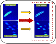 .
.
CONFLICT OF INTEREST
The authors declare no financial or commercial conflict of interest.
Open Research
DATA AVAILABILITY STATEMENT
Research data are not shared.