Volume 13, Issue 10 e202000212
LETTER

Deep learning protocol for improved photoacoustic brain imaging

Rayyan Manwar

Rayyan Manwar

Richard and Loan Hill Department of Bioengineering, University of Illinois at Chicago, Chicago, Illinois, USA

Department of Biomedical Engineering, Wayne State University, Detroit, Michigan, USA

Search for more papers by this author
Xin Li

Xin Li

Department of Computer Science, Wayne State University, Detroit, Michigan, USA

Search for more papers by this author
Sadreddin Mahmoodkalayeh

Sadreddin Mahmoodkalayeh

Department of Physics, Shahid Beheshti University, Tehran, Iran

Search for more papers by this author
Eishi Asano

Eishi Asano

Departments of Pediatrics and Neurology, School of Medicine, Wayne State University, Detroit, Michigan, USA

Search for more papers by this author
Dongxiao Zhu

Dongxiao Zhu

Department of Computer Science, Wayne State University, Detroit, Michigan, USA

Search for more papers by this author
Kamran Avanaki

Corresponding Author

Kamran Avanaki

Richard and Loan Hill Department of Bioengineering, University of Illinois at Chicago, Chicago, Illinois, USA

Department of Biomedical Engineering, Wayne State University, Detroit, Michigan, USA

Correspondence

Kamran Avanaki, Richard and Loan Hill Department of Bioengineering, University of Illinois at Chicago, Chicago, IL, 60607; Department of Biomedical Engineering, Wayne State University, Detroit, MI.

Email: [email protected]

Search for more papers by this author
First published: 21 July 2020
Citations: 48

Funding information: National Institutes of Health, Grant/Award Numbers: R01EB027769-01, R01EB028661-01

Abstract

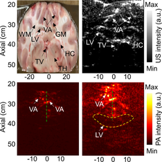
One of the key limitations for the clinical translation of photoacoustic imaging is penetration depth that is linked to the tissue maximum permissible exposures (MPE) recommended by the American National Standards Institute (ANSI). Here, we propose a method based on deep learning to virtually increase the MPE in order to enhance the signal-to-noise ratio of deep structures in the brain tissue. The proposed method is evaluated in an in vivo sheep brain imaging experiment. We believe this method can facilitate clinical translation of photoacoustic technique in brain imaging, especially in transfontanelle brain imaging in neonates.image

The full text of this article hosted at iucr.org is unavailable due to technical difficulties.