

Raman microspectroscopic study for the detection of oral field cancerisation using brush biopsy samples
Corresponding Author
Isha Behl
Centre for Radiation and Environmental Science, FOCAS Research Institute, Technological University Dublin, Dublin, Ireland
School of Physics, Technological University Dublin, Dublin, Ireland
Correspondence
Dr Isha Behl, Centre for Radiation and Environmental Science, FOCAS Research Institute, Technological University Dublin, City Campus, Dublin, Ireland.
Email: [email protected]
Search for more papers by this authorGenecy Calado
Centre for Radiation and Environmental Science, FOCAS Research Institute, Technological University Dublin, Dublin, Ireland
School of Physics, Technological University Dublin, Dublin, Ireland
Search for more papers by this authorAnika Vishwakarma
Centre for Radiation and Environmental Science, FOCAS Research Institute, Technological University Dublin, Dublin, Ireland
School of Physics, Technological University Dublin, Dublin, Ireland
Search for more papers by this authorStephen Flint
Oral Medicine Unit, Dublin Dental University Hospital, Trinity College Dublin, Dublin, Ireland
Search for more papers by this authorSheila Galvin
Oral Medicine Unit, Dublin Dental University Hospital, Trinity College Dublin, Dublin, Ireland
Search for more papers by this authorClaire M. Healy
Oral Medicine Unit, Dublin Dental University Hospital, Trinity College Dublin, Dublin, Ireland
Search for more papers by this authorMarina Leite Pimentel
Division of Restorative Dentistry and Periodontology, Dublin Dental University Hospital, Trinity College Dublin, Dublin, Ireland
Search for more papers by this authorAlison Malkin
School of Biological Sciences, Technological University Dublin, Dublin, Ireland
Search for more papers by this authorHugh J. Byrne
FOCAS Research Institute, Technological University Dublin, Dublin, Ireland
Search for more papers by this authorFiona M. Lyng
Centre for Radiation and Environmental Science, FOCAS Research Institute, Technological University Dublin, Dublin, Ireland
School of Physics, Technological University Dublin, Dublin, Ireland
Search for more papers by this authorCorresponding Author
Isha Behl
Centre for Radiation and Environmental Science, FOCAS Research Institute, Technological University Dublin, Dublin, Ireland
School of Physics, Technological University Dublin, Dublin, Ireland
Correspondence
Dr Isha Behl, Centre for Radiation and Environmental Science, FOCAS Research Institute, Technological University Dublin, City Campus, Dublin, Ireland.
Email: [email protected]
Search for more papers by this authorGenecy Calado
Centre for Radiation and Environmental Science, FOCAS Research Institute, Technological University Dublin, Dublin, Ireland
School of Physics, Technological University Dublin, Dublin, Ireland
Search for more papers by this authorAnika Vishwakarma
Centre for Radiation and Environmental Science, FOCAS Research Institute, Technological University Dublin, Dublin, Ireland
School of Physics, Technological University Dublin, Dublin, Ireland
Search for more papers by this authorStephen Flint
Oral Medicine Unit, Dublin Dental University Hospital, Trinity College Dublin, Dublin, Ireland
Search for more papers by this authorSheila Galvin
Oral Medicine Unit, Dublin Dental University Hospital, Trinity College Dublin, Dublin, Ireland
Search for more papers by this authorClaire M. Healy
Oral Medicine Unit, Dublin Dental University Hospital, Trinity College Dublin, Dublin, Ireland
Search for more papers by this authorMarina Leite Pimentel
Division of Restorative Dentistry and Periodontology, Dublin Dental University Hospital, Trinity College Dublin, Dublin, Ireland
Search for more papers by this authorAlison Malkin
School of Biological Sciences, Technological University Dublin, Dublin, Ireland
Search for more papers by this authorHugh J. Byrne
FOCAS Research Institute, Technological University Dublin, Dublin, Ireland
Search for more papers by this authorFiona M. Lyng
Centre for Radiation and Environmental Science, FOCAS Research Institute, Technological University Dublin, Dublin, Ireland
School of Physics, Technological University Dublin, Dublin, Ireland
Search for more papers by this authorFunding information: Science Foundation Ireland, Grant/Award Number: 12/IP/1494; Technological University Dublin, Fiosraigh Dean of Graduate Studies Award
Abstract
Field cancerisation (FC) is potentially an underlying cause of poor treatment outcomes of oral squamous cell carcinoma (OSCC). To explore the phenomenon using Raman microspectroscopy, brush biopsies from the buccal mucosa, tongue, gingiva and alveolus of healthy donors (n = 40) and from potentially malignant lesions (PML) of Dysplasia Clinic patients (n = 40) were examined. Contralateral normal samples (n = 38) were also collected from the patients. Raman spectra were acquired from the nucleus and cytoplasm of each cell, and subjected to partial least squares-discriminant analysis (PLS-DA). High discriminatory accuracy for donor and PML samples was achieved for both cytopalmic and nuclear data sets. Notably, contralateral normal (patient) samples were also accurately discriminated from donor samples and contralateral normal samples from patients with multiple lesions showed a similar spectral profile to PML samples, strongly indicating a FC effect. These findings support the potential of Raman microspectroscopy as a screening tool for PML using oral exfoliated cells.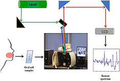
CONFLICT OF INTEREST
The authors declare no potential conflict of interest.
Supporting Information
| Filename | Description |
|---|---|
| jbio202000131-sup-0001-Figure S1.jpgJPEG image, 469 KB | Figure S1 a) Score plot for healthy volunteer samples and patient (potentially malignant lesion [lesion]) samples from cytoplasm of gingiva and alveolus, b) Latent variable loading, c) Probability predictive plot, d) Confusion matrix |
| jbio202000131-sup-0002-Figure S2.jpgJPEG image, 512.3 KB | Figure S2 a) Score plot for healthy volunteer samples and patient (potentially malignant lesion [lesion]) samples from nucleus of gingiva and alveolus, b) Latent variable loading, c) Probability predictive plot, d) Confusion matrix |
| jbio202000131-sup-0003-Figure S3.jpgJPEG image, 446.4 KB | Figure S3 a) Score plot for healthy volunteer samples and patient (contralateral normal) samples from cytoplasm of gingiva and alveolus, b) Latent variable loading, c) Probability predictive plot, d) Confusion matrix |
| jbio202000131-sup-0004-Figure S4.jpgJPEG image, 454.9 KB | Figure S4 a) Score plot for healthy volunteer samples and patient (contralateral normal) samples from nucleus of gingiva and alveolus, b) Latent variable loading, c) Probability predictive plot, d) Confusion matrix |
| jbio202000131-sup-0005-Figure S5.jpgJPEG image, 492.3 KB | Figure S5 a) Score plot for potentially malignant lesion (lesion) samples and contralateral normal samples from cytoplasm of gingiva and alveolus, b) Latent variable loading, c) Probability predictive plot, d) Confusion matrix |
| jbio202000131-sup-0006-Figure S6.jpgJPEG image, 479.3 KB | Figure S6 a) Score plot for potentially malignant lesion (lesion) samples and contralateral normal samples from nucleus of gingiva and alveolus, b) Latent variable loading, c) Probability predictive plot, d) Confusion matrix |
Please note: The publisher is not responsible for the content or functionality of any supporting information supplied by the authors. Any queries (other than missing content) should be directed to the corresponding author for the article.
REFERENCES
- 1GLOBOCAN: Estimated Cancer Incidence, Mortality and Prevalence Worldwide in 2018. International Agency for Research on Cancer [online] 2018, http://globocan.iarc.fr/Pages/fact_sheets_population.aspx.
- 2B. W. Neville, T. Day, Ca-A Cancer J. Clin. 2002, 52(4), 195.
- 3A. C. Sathiasekar, D. G. Mathew, M. S. Jaish Lal, A. A. Arul Prakash, K. U. G. Kumar, J Pharm Bioallied Sci. 2017, 9(Suppl 1), S23.
- 4T. A. Winning, G. C. Townsend, Clin. Dermatol. 2000, 18(5), 499.
- 5C. A. Squier, M. J. Kremer, J. Natl. Cancer Inst. Monogr. 2001, 52242(29), 7.
10.1093/oxfordjournals.jncimonographs.a003443 Google Scholar
- 6V. Kumar, N. Fausto, A. Abbas, Robbins and Cotran Pathologic Basis of Disease, 7th ed., Elsevier, Amsterdam, 2005.
- 7 A. K. El-Naggar, J. K. C. Chan, J. R. Grandis, T. Takata, P. J. Slootweg Eds., WHO Classification of Head and Neck Tumours, 4th ed., IARC, France, 2017.
- 8S. Müller, Oral Surg. Oral Med. Oral Pathol. Oral Radiol. 2018, 125(6), 591.
- 9C. F. Poh, C. E. MacAulay, D. M. Laronde, P. M. Williams, L. Zhang, M. P. Rosin, Periodontol 2000. 2011, 57(1), 73.
- 10D. P. Slaughter, H. W. Southwick, W. Smejkal, Cancer 1953, 6, 963.
10.1002/1097-0142(195309)6:5<963::AID-CNCR2820060515>3.0.CO;2-Q CAS PubMed Web of Science® Google Scholar
- 11M. Mohan, N. Jagannathan, Oncol. Rev. 2014, 8(1), 244. Doi: 10.4081/oncol.2014.244.
- 12M. Aparna, P. Shenai, L. K. Chatra, K. M. Veena, P. K. Rao, R. V. Prabhu, K. A. Shahin, Arch. Med. Heal. Sci. 2013, 1(2), 136.
10.4103/2321-4848.123026 Google Scholar
- 13M. G. C. T. van Oijen, P. J. Slootweg, Cancer Epidemiol. Biomarkers Prev. 2000, 9(3), 249.
- 14S. P. Singh, A. Deshmukh, P. Chaturvedi, C. M. Krishna, J. Biomed. Opt. 2012, 17(10), 1050021.
- 15A. Sahu, S. Tawde, V. Pai, P. Gera, P. Chaturvedi, S. Nair, C. M. Krishna, Anal. Methods 2015, 7, 7548.
- 16A. Sahu, N. Shah, M. Mahimkar, M. Garud, S. Pagare, S. Nair, C. M. Krishna, in Proc. SPIE 8926, Photon Therap Diagn X, 89262N, 2014.
- 17A. Sahu, A. Deshmukh, A. D. Ghanate, S. P. Singh, P. Chaturvedi, C. M. Krishna, Technol. Cancer Res. Treat. 2012, 11, 529.
- 18S. P. Singh, PhD, Tata Memorial Centre 2013.
- 19S. P. Singh, A. Deshmukh, P. Chaturvedi, C. M. Krishna, J. Cancer Res. Ther. 2012, 8(6), 126.
- 20I. Behl, L. Kukreja, A. Deshmukh, S. P. Singh, H. Mamgain, A. R. Hole, C. M. Krishna, J. Biomed. Opt. 2014, 19(12), 126005.
- 21H. Krishna, S. K. Majumder, P. Chaturvedi, M. Sidramesh, P. K. Gupta, J. Biophotonics 2014, 7(9), 690.
- 22M. J. Jeng, M. Sharma, L. Sharma, T. Y. Chao, S. F. Huang, L. B. Chang, S. L. Wu, L. Chow, J. Clin. Med. 2019, 8(9), 1313.
- 23F. L. J. Cals, T. C. Bakker Schut, P. J. Caspers, R. J. Baatenburg De Jong, S. Koljenović, G. J. Puppels, Analyst 2018, 143(17), 4090.
- 24C. LFCS, F. Bonnier, C. Tellez, L. Dos Santos, K. O'Callaghan, J. O'Sullivan, S. LES, S. Flint, A. A. Martin, F. M. Lyng, H. J. Byrne, Exp. Mol. Pathol. 2017, 103(3), 255.
- 25F. L. Cals, T. C. Bakker Schut, J. A. Hardillo, R. J. Baatenburg de Jong, S. Koljenović, G. J. Puppels, Lab. Invest. 2015, 95(10), 1186.
- 26L. F. C. S. Carvalho, F. Bonnier, K. O. Callaghan, J. O. Sullivan, S. Flint, H. J. Byrne, F. M. Lyng, Exp. Mol. Pathol. 2015, 98(3), 502.
- 27L. F. C. S. Carvalho, M. S. Nogueira, T. Bhattacharjee, L. P. M. Neto, L. Daun, T. O. Mendes, R. Rajasekaran, M. Chagas, A. A. Martin, L. E. S. Soares, Clin. Oral Investig. 2019, 23(7), 3021.
- 28L. F. C. S. Carvalho, M5. S. Nogueira, Analyst 2018, 143(24), 6037.
- 29C. Wu, J. Gleysteen, N. T. Teraphongphom, Y. Li, E. Rosenthal, Int. J. Oral Sci. 2018, 10(2), 10.
- 30C. A. Lieber, H. E. Nethercott, M. H. Kabeer, Biomed. Opt. Express 2010, 1(3), 975.
- 31A. Robichaux Viehoever, D. Anderson, D. Jansen, A. Mahadevan-Jansen, Photochem. Photobiol. 2004, 78(5), 517.
- 32S. P. Singh, A. Sahu, A. Deshmukh, P. Chaturvedi, C. M. Krishna, Analyst 2013, 138, 4175.
- 33I. Behl, G. Calado, O. Ibrahim, A. Malkin, S. Flint, H. J. Byrne, F. M. Lyng, Anal. Methods 2017, 9(6), 937.
- 34F. Bonnier, D. Traynor, P. Kearney, C. Clarke, P. Knief, C. Martin, J. J. O'Leary, H. J. Byrne, F. M. Lyng, Anal. Methods 2014, 6, 7831.
- 35L. T. Kerr, B. M. Hennelly, Chemom. Intel. Lab. Syst. 2016, 158, 61.
- 36M. Urvoy, F. Autrusseau, in Proc. 2nd ACM Work. Inf. Hiding Multimed. Secur. - IH&MMSec '14, 2014, pp. 49–60.
- 37R. G. Brereton, G. R. Lloyd, J. Chemometr. 2014, 28(4), 213.
- 38R. Gautam, S. Vanga, F. Ariese, S. Umapathy, EPJ Tech. Instrum. 2015, 2(1), 8.
- 39I. Behl, G. Calado, A. Malkin, S. Flint, S. Galvin, C. M. Healy, M. L. Pimentel, H. J. Byrne, F. M. Lyng, J. Biophotonics 2020, https://doi.org/10.1002/jbio.202000079.
- 40Z. Movasaghi, S. Rehman, I. Rehman, Appl. Spectrosc. 2007, 42(5), 493.
- 41H. Byrne, G. Sockalingum, N. Stone, Raman Microscopy: Complement or Competitor. in Biomedical Applications of Synchrotron Infrared Microspectroscopy, RSC Analytical Spectroscopy Series 2011, p. 105-143.
- 42S. Beloribi-Djefaflia, S. Vasseur, F. Guillaumond, Oncogenesis 2016, 5(1), e189.
- 43G. Cascianelli, M. Villani, M. Tosti, F. Marini, E. Bartoccini, M. V. Magni, E. Albi, Mol. Biol. Cell 2008, 19(12), 5289. https://doi.org/10.1091/mbc.e08-05-0517.
- 44I Behl, PhD, TU Dublin, 2019.
- 45D. V. Dawson, D. R. Drake, J. R. Hill, K. A. Brogden, C. L. Fischer, P. W. Wertz, Int. J. Cosmet. Sci. 2013, 35(3), 220.
- 46R. J. DeBerardinis, A. Mancuso, E. Daikhin, I. Nissim, M. Yudkoff, S. Wehrli, C. B. Thompson, Proc. Natl. Acad. Sci. USA 2007, 104(49), 19345.
- 47E. L. Lieu, T. Nguyen, S. Rhyne, J. Kim, Exp. Mol. Med. 2020, 52(1), 15.
- 48R. J. DeBerardinis, S. C. Navdeep, Oncology 2016, 2, 1.
- 49L. M. Phan, S. C. J. Yeung, M. H. Lee, Cancer Biol. Med. 2014, 11(1), 1.
- 50J. Giovannoni, M. G. C. T. Van Oijen, P. J. Slootweg, M. Mohan, N. Jagannathan, Oncol. Rev. 2014, 8(1), 249.
Citing Literature
