Rapid intraoperative diagnosis of gynecological cancer by ATR-FTIR spectroscopy of fresh tissue biopsy
Funding information: Israel Cancer Association (ICA), Grant/Award Number: 20191636
Abstract
A rapid and reliable intraoperative diagnostic technique to support clinical decisions was developed using Fourier-transform infrared (FTIR) spectroscopy. Twenty-six fresh tissue samples were collected intraoperatively from patients undergoing gynecological surgeries. Frozen section (FS) histopathology aimed to discriminate between malignant and benign tumors was performed, and attenuated total reflection (ATR) FTIR spectra were collected from these samples. Digital dehydration and principal component analysis and linear discriminant analysis (PCA-LDA) models were developed to classify samples into malignant and benign groups. Two validation schemes were employed: k-fold and “leave one out.” FTIR absorption spectrum of a fresh tissue sample was obtained in less than 5 minutes. The fingerprint spectral region of malignant tumors was consistently different from that of benign tumors. The PCA-LDA discrimination model correctly classified the samples into malignant and benign groups with accuracies of 96% and 93% for the k-fold and “leave one out” validation schemes, respectively. We showed that a simple tissue preparation followed by ATR-FTIR spectroscopy provides accurate means for very rapid tumor classification into malignant and benign gynecological tumors. With further development, the proposed method has high potential to be used as an adjunct to the intraoperative FS histopathology technique.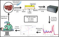
CONFLICT OF INTEREST
The authors declare that they have no conflict of interest.
Open Research
DATA AVAILABILITY STATEMENT
The data that support the findings of this study are available from the corresponding author upon request.