Cover Picture
Free Access
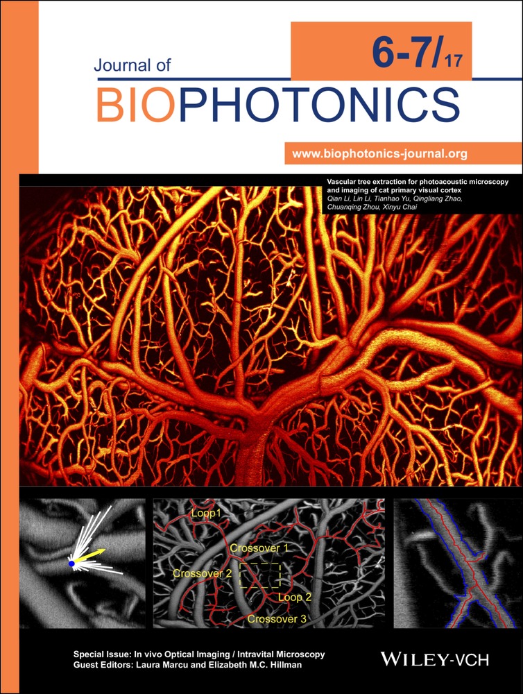
Front Cover: Vascular tree extraction for photoacoustic microscopy and imaging of cat primary visual cortex (J. Biophotonics 6-7/2017)
First published: 29 June 2017
Graphical Abstract
Photoacoustic imaging allows label-free, three-dimensional visualization of blood vessel morphology down to capillary level. By virtue of the acquired high contrast blood vessel images, a vascular tree extraction method based on the ray-casting concept could automatically track complex cortical blood vessel networks of cats with numerous crossovers and branches, while extracting important parameters of the whole vascular tree, such as the vessel diameters, the center lines and three-dimensional orientations. Further details can be found in the article by Qian Li et al. on pp. 780–791.