Volume 10, Issue 9 e6380
CASE IMAGE
Open Access

An unusual simultaneous occurrence of gastric carcinoma with lymphoid stroma, calcified leiomyoma and ectopic pancreas

Faten Limaiem

Corresponding Author

Faten Limaiem

Tunis Faculty of Medicine, University of Tunis El Manar, Tunis, Tunisia

Pathology Department, University Hospital Mongi Slim, Tunis, Tunisia

Correspondence

Faten Limaiem, Department of Pathology, Mongi Slim Hospital, La Marsa, Tunisia.

Email: [email protected]

Search for more papers by this author
Sahir Omrani

Sahir Omrani

Tunis Faculty of Medicine, University of Tunis El Manar, Tunis, Tunisia

General Surgery Department, University Hospital Mongi Slim, Tunis, Tunisia

Search for more papers by this author
First published: 25 September 2022

Abstract

Gastric carcinoma with lymphoid stroma is a rare variant of gastric carcinoma accounting for 1%–7% of gastric carcinomas. Its association with gastric leiomyoma and ectopic pancreas is extremely rare. We herein report an unusual simultaneous occurrence of gastric carcinoma with lymphoid stroma, calcified leiomyoma, and ectopic pancreas.

1 CLINICAL IMAGE

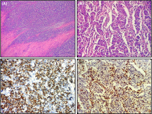
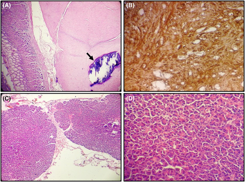
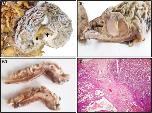
A 65-year-old woman with a past medical history of breast cancer and hypertension, presented with a 3-month history of epigastralgia. Upper gastrointestinal endoscopy showed a vegetative mass in the antrum. Histological examination of the biopsy specimens revealed gastric adenocarcinoma. The patient underwent subtotal gastrectomy. Gross examination of the surgical specimen disclosed two contiguous mass lesions located in the antrum (Figure 1A,B). The first tumor was polypoid sharply demarcated from the surrounding mucosa. The second tumor occupied the anterior and posterior wall of the stomach. In the fundus, there was a 2-cm submucosal tumor (Figure 1C). Microscopic examination of the surgical specimen showed the coexistence of two tumor components including gastric carcinoma with lymphoid stroma and tubular adenocarcinoma (Figures 1D and 2A,B). Immunohistochemical study showed positive immunostaining of tumor cells with cytokeratin (Figure 2C). The prominent lymphocytic infiltrate of the stroma was CD3 positive (Figure 2D). The 2-cm submucosal lesion corresponded to a calcified leiomyoma (Figure 3A) which showed positive immunostaining with SMA and desmin (Figure 3B).1, 2 We also incidentally discovered the presence of a tiny foci of ectopic pancreas within the greater omentum intermixed with adipose tissue (Figure 3C,D). On postoperative day 7, the patient died due to perianastomotic abscess and peritonitis.
Details are in the caption following the image
(A) Subtotal gastrectomy specimen showing two contiguous mass lesions located in the antrum. The first tumor is polypoid sharply demarcated from the surrounding mucosa (yellow asterisk). The second tumor contiguous to the first infiltrated the wall of the antrum. It was white in color and occupied the anterior and posterior wall of the stomach (black arrows). (B) Cut section of the tumor showing a polypoid and fungating tumor with broad base (yellow asterisk). The surrounding gastric mucosa was abnormally thickened (white asterisk). (C) Macroscopic findings of gastric leiomyoma. On the cut surface, we note the presence of a firm whitish submucosal nodule measuring 2 cm in diameter (blue asterisk). (D) Representative photomicrograph of gastric carcinoma with two compartments. The first compartment (at the right of the figure) is gastric carcinoma with lymphoid stroma. The second component (at the left of the figure) shows glandular differentiation (Hematoxylin and eosin, magnification ×40).
Details are in the caption following the image
(A) Gastric carcinoma with lymphoid stroma infiltrating the muscularis. Irregular sheets and trabeculae of polygonal cells embedded within a prominent lymphocytic infiltrate (Hematoxylin and eosin, magnification ×100). (B) Gastric carcinoma with lymphoid stroma. Neoplastic cells are arranged in small nests or trabeculae accompanied by an abundant lymphocyte infiltration (Hematoxylin and eosin, magnification ×400). (C) Immunohistochemistry demonstrating strong and diffuse staining of the tumor cells with cytokeratin (Immunohistochemistry, magnification ×400). (D) Immunohistochemistry demonstrating staining of the lymphoid cells with CD3 (brown staining) (Immunohistochemistry, magnification ×400).
Details are in the caption following the image
(A) Histological findings of a submucosal calcified leiomyoma. In the submucosa of the fundus, we note the presence of bundles of interlacing smooth muscle cells. Within the tumor proliferation we note the presence of calcification (black arrow) (Hematoxylin and eosin, magnification ×40). (B) Immunohistochemistry demonstrating strong and diffuse staining of the tumor cells with smooth muscle actin (SMA) (brown staining) (Immunohistochemistry, magnification ×400). (C) Histological findings of the heterotopic pancreas. Within the adipose tissue of the greater omentum, there were lobules made of benign-appearing pancreatic acini and ducts (Hematoxylin and eosin, magnification ×100). (D) Histological findings of heterotopic pancreas made of benign-appearing pancreatic acini (Hematoxylin and eosin, magnification ×400).

AUTHOR CONTRIBUTIONS

Faten Limaiem prepared, organized, wrote, and edited all aspects of the manuscript and prepared all of the histology figures in the manuscript. Sahir Omrani participated in the conception and design of the study, the acquisition of data, analysis and interpretation of the data. Both authors read, edited, and approved the final version of the manuscript. They contributed equally to preparing the manuscript and participated in the final approval of the manuscript before its submission.

ACKNOWLEDGMENTS

None.

    CONFLICT OF INTEREST

    None declared.

    ETHICAL APPROVAL

    All procedures performed were in accordance with the ethical standards. The examination was made in accordance with the approved principles.

    CONSENT

    Published with written consent of the patient.

    DATA AVAILABILITY STATEMENT

    The data that support the findings of this study are available from the corresponding author upon reasonable request.

      The full text of this article hosted at iucr.org is unavailable due to technical difficulties.