Severe mucosal leishmaniasis with torpid and fatal evolution
Abstract
Mucosal leishmaniasis is a clinical condition that is difficult to diagnose and treat and usually precedes a cutaneous leishmaniasis condition with a long latency period as observed in our study of a patient who experienced a torpid evolution in 9 months, caused by having had cutaneous leishmaniasis on the neck without therapeutic treatment, although with ulcer closure 18 years earlier, incomplete treatment with antimonials and amphotericin B, with the destruction of the eyeball, a large area of necrosis on the face and nasal bone exposure. Additionally, the patient had chronic anemia (9.4 g/dl), lymphopenia and neutrophilia (lymphocytes 13.1%, neutrophils 84.4%), and co-infections by fungi (yeasts and hyphae) and Gram-negative bacteria (multidrug-resistant Proteus mirabilis and Escherichia coli) leading to sepsis and subsequent death of the patient.
1 INTRODUCTION
Leishmaniasis is a disease caused by the protozoan Leishmania sp. and its 22 pathogenic species. This parasite is transmitted by the vector Lutzomyia sp. in the New World with the clinical form and drug resistance depending on the species-host relationship.1, 2 The disease in the America may present three classic clinical forms: cutaneous, mucosal, and visceral3 although there are also intermediate clinical forms such as mucocutaneous, which is one of the most severe forms of the disease.4 In some cases, the mucosal lesions can be extensive in centimeters and may include areas of the face or other mucous membranes.5, 6 Mucosal lesions are a chronic degenerative phase that usually appears more than two years after the patient has presented a skin lesion.7 In addition, it is known that in cases of mucosal leishmaniasis (ML) destructive lesions are irreversible leading to respiratory complications and malnutrition, and between 3% and 5% of cases of cutaneous leishmaniasis develop into mucosal leishmaniasis with tissue destruction in future years.8, 9
Patient nutrition is a key factor in disease remission and is related to all physiological processes including the immune system.5 Malnutrition results in the exacerbation of neglected diseases including leishmaniasis.10 The relationship between malnutrition and mucosal leishmaniasis has been documented with a special relationship in the depletion of T lymphocytes that play a key role in the cure or progression of the disease.11, 12
In addition, it is known that the parasite can live for a long time in the organism13 even in cases where antimonial-based therapy has been administered, complicating the diagnosis and therapy to be implemented. And among the parasitic species mainly attributed to LM are Leishmania braziliensis and Leishmania guyanensis.14
This study aimed to describe an unusual case of a patient with mucosal leishmaniasis with a fatal ending.
2 CASE REPORT
Data were collected on the patient's age, gender, and residence during the illness. The presence of the Leishmania parasite was determined by Indirect Immunofluorescence (IFA), histology, kDNA-PCR, and Nested-PCR. In addition, serological tests for HIV, hemogram, and multislice spiral tomography were taken. The use of antileishmanicidal drugs (sodium stibogluconate, Glucantime, and amphotericin B), their dosage, and duration during the period of the disease were reported. The clinical course of the patient's illness from the onset of the first cutaneous infection to the development of the mucosal episode was reported. Aggregate coinfections and the patient's prognosis were also described. Immunocompetent male patient with no comorbidities, 44 years old, 64.5 kg, and 1.55 m tall, resident of Puerto Maldonado, Madre de Dios-Peru, who worked in artisanal gold mining, timber, and chestnut extraction.
When he was 24-year-old, he developed a clinical condition compatible with the diagnosis of cutaneous leishmaniasis on the neck and with apparent total healing of the ulcer without using antimonial treatment. At the age of 34 years, he presented 14% of eosinophils with a leukocyte count of 10,200/mm3. Parasitological diagnosis identified eggs of Uncinaria sp. (+) in the feces which was not reported to have been treated. When the patient was 42-year-old, he reported slight nasal bleeding and after 3 months, he began to drain a fetid yellow liquid.
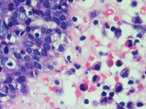
The case of this disease began as a nasal tumor which the patient, 9 months before his death, was diagnosed in the city of Arequipa by histology, in which an irregular tissue of 2.8 cm in diameter was obtained from the left nasal turbinate where lymphoplasmacytic infiltrate, fibrosis, and extensive areas with necrosis and presence of Leishmania sp. amastigotes were observed (Figure 1). Indirect immunofluorescence was negative, hemoglobin of 13.90 gr/dl, and 16,630 leukocytes/mm3.

Subsequently, he returned to Puerto Maldonado—Madre de Dios and received intravenous sodium stibogluconate for 28 days, at a dose of 20 mg/kg, with a maximum dose of 1200 mg/day, at the Jorge Chávez Health Center. At the end of treatment, a muco-nasal lesion was identified. After 15 days, the patient received meglumine antimoniate “Glucantime” in doses of 2 ampoules (1500 mg/ 5 ml) per day as an outpatient without determining if the treatment was daily for approximately 2 months, after which it was observed that the lesion did not remit.
Then, the patient traveled to the city of Cusco, to the regional hospital where he was administered amphotericin B 30 mg/24 h x 25 days, vancomycin 500 mg/8 h, ceftazidime 1 gr/8 h, and ciprofloxacin 200 mg/12 h. Complementary, laboratory tests for HIV ELISA were performed and were negative. Multislice spiral tomography (CT) of the paranasal sinuses without contrast showed abundant liquid in the paranasal sinuses and ethmoidal cells, morphological distortion of both ostiomeatal complexes, and deviation of the bony nasal septum to the right, concluding in pansinusitis without remission of the nasal lesion. The patient asked for his voluntary discharge due to the side effects of amphotericin B.
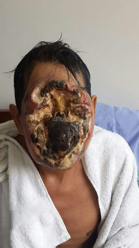
Five months after the onset of the clinic the patient returns to Puerto Maldonado-Madre de Dios and remains untreated for 3 months and the lesion spreads over the face. On admission to the Santa Rosa Hospital in Puerto Maldonado (25/08/15), a disease period of 9 months is mentioned, infection of extensive deep facial lesion of 18 × 15 cm from the frontal region to the upper jaw, irregular erythematous edges with the presence of purulent fluid with areas of necrosis with exposure of nasal bone and destruction of the eyeball involving both eyes and nose was observed (Figure 2). The therapeutic regimen was intravenous ceftazidime 1 gr/8 h, intravenous vancomycin 1 gr/12 h, paracetamol 750 mg/8 h, and intravenous ranitidine 50 mg/8 h. The condition presents chronic malnutrition and the absence of teeth. In the opinion of surgery, surgical cleaning is not possible due to the possibility of bleeding. Laboratory tests showed 9.4 gr/dl of hemoglobin, lymphocytes (13.1%), monocytes (2.2%), eosinophils (0.3%) concluding in lymphopenia and a high value of neutrophils (84.4%). The histological result of nasal tissue biopsy was positive for Leishmania, as well as the kDNA-PCR molecular test was positive for Leishmania. Subsequently, a real-time Nested PCR was performed to identify the parasite species, but it was negative due to low parasitemia. The microbiological examination of facial secretions revealed Proteus mirabilis resistant to sulfatrimetropin (1.25/23.75 μg), gentamicin (10 μg), ciprofloxacin (5 μg), and clindamycin (10 μg), Escherichia coli resistant to sulfatrimetropin (1.25/23.75 μg), gentamicin (10 μg), cefuroxime (30 μg), ciprofloxacin (5 μg), amoxicillin/clavulanic acid (20/10 μg), cefotaxime (30 μg), and clindamycin (10 μg), as well as Gram-positive bacteria (+), yeasts (++), and hyphae (+).

The patient's relative (wife) does not want her husband to be referred to a more complex hospital in Lima because she says that she does not have the financial resources to support herself in the capital and she needs to take care of her young children. The day after his admission, he presented dysphagia and difficulty in ingesting food, without fever. Three days after his admission, the wounds showed profuse bleeding from the orbital holes; no family members were present, the disease worsened with a torpid evolution and the patient showed septicemia, malnutrition, and depression, dying of septic shock.
3 DISCUSSION
Mucosal leishmaniasis is the most serious tegumentary disease in relation to sequelae and co-infections, being difficult to diagnose and treat, and usually destroys areas of the respiratory cavity in which necrotic sections are observed, as the case of our patient in the present study.15 Although the presence of Leishmania sp. was confirmed by histology and PCR-kinetoplast, the species was not detected. In cases of mucosal leishmaniasis, low parasite load has been reported16 as observed by histology and PCR-kinetoplast band in our study.
The patient had a case of cutaneous leishmaniasis on the neck 20 years before the onset of the case of severe mucosal leishmaniasis. Although the primary lesion of 20 years ago healed, studies have shown that the parasites can remain in the tissue for a long time besides the inflammatory process, so it is recurrent. In some cases, a secondary lesion can be developed after many years, even in cases where antimonials were used.17, 18 Leishmaniasis is endemic in the region of Madre de Dios in Peru, where one of the most prevalent species is L. braziliensis, the main causal agent of mucocutaneous and mucosal leishmaniasis.12, 19, 20
L. braziliensis is related to the development of secondary disease with long latency periods after the first lesion. A previous case showed a latency period of almost 30 years after the primary lesion caused by L. braziliensis.9, 21 There are known cases of patients who develop primary cutaneous leishmaniasis and several years later develop mucosal leishmaniasis caused mainly by L. braziliensis in a process that causes chronic inflammation and hyperreactivity of CD8+ T-cell response with high levels of TNFα, ITFϒ, and IL-2, and decreased IL-10 and synthesis of granzymes and perforins with high cytolytic activity resulting in the recruitment of monocytes and neutrophils that secrete IL-1 β, which generates the pathophysiology and severity of the disease. TCD8+ can also secrete IL-17 which is associated with infiltration and recruitment of neutrophils in the lesion, promoting a cyclic inflammatory response with production of nitric oxide (NO) which plays a dual role in parasite elimination and inflammatory response.12, 22, 23 Highlighting that the constantly secreted proinflammatory cytokines (IL-2 and IL-17) are associated with the destruction of tissues causing coinfections that stimulate greater secretion of proinflammatory cytokines in a cyclical phenomenon.
In our case, a potent inflammatory response was observed in relation to neutrophilia contributing to tissue damage besides a marked lymphocytopenia. The uncontrolled and non-specific inflammatory process is characteristic of mucosal leishmaniasis.12 In these cases, the injury is exacerbated by an inflammatory process that may last for months or even years despite the decrease in parasite load triggered by inflammatory cytokines secreted in the cell response and where there may be dysfunction of the Th1/Th2 balance.17, 24, 25 Chronic granulomatosis with a chronic inflammatory process and lymphocytoplasmacytic infiltration characteristic of mucosal leishmaniasis was observed in the tissue at the onset of the disease, as reported in a previous study.26
The patient who had an evolution of mucosal leishmaniasis over 9 months initially received the antimonial (sodium stibogluconate) treatment incompletely, as therapy at 20 mg/Kg/day (only 28 days), since the protocol in case of mucosal leishmaniasis indicates an initial treatment of 30 days.27 Subsequently, he received Glucantime for 2 months as an informal outpatient, as well as homemade plant-based plasters for 3 months and amphotericin B (25 days). The therapeutic failure is related to incomplete dosage, the use of substances without proven antiparasitic effect as homemade remedies that could exacerbate the disease generating the pathophysiology mediated by proinflammatory cytokines with areas of necrosis,12, 22, 23 as observed histologically and clinically.
Incomplete treatment with antimonials is a risk factor for disease progression,6 because, although it stimulates the synthesis of nitric oxide as an antiparasitic, incomplete treatment with antimonials provides the opportunity to maintain the parasite load that promotes the recruitment of nuclear polymorphs, exacerbating the inflammatory process stimulated by the presence of TNF α, IL-1, and nitric oxide due to the presence of the parasite and germs that co-infect the injured area in mucosal leishmaniasis,12 as it was determined in the patient.
Antimonials are drugs that cause serious adverse effects including aplastic anemia, renal and hepatic impairment, so, he should have reported these effects, and, additionally, modest therapeutic rates are reported with this drug.28 Although the clinical form depends on the infecting Leishmania species and the patient's immune response,29 L. braziliensis is a species related to high resistance to antimonials used as therapy, so its persistence in the body leads to a chronic inflammatory state.2 Amphotericin B was also used at 0.5 mg/kg/day up to a maximum of 50 mg/day, although the patient received incomplete therapy (only 25 days out of the recommended 60 days).27 According to the report, the patient abandoned the treatment because of the side effects such as severe headaches and nausea due to the drug, as previously reported in an earlier study.30
Additionally, the patient's hemoglobin at the beginning of the diagnosis of mucosal leishmaniasis was 13.90 g/dl, very close to the cut-off point for diagnosing the patient with anemia, and 9 months later it dropped to 9.4 g/dl with a diagnosis of chronic anemia, a marked lymphocytopenia and neutrophilia diagnosed in the final stage of the disease, where the lymphocytopenia observed in the patient could indicate a decrease of IL-10, increasing the number of proinflammatory cytokines with antiparasitic effect, although mediating tissue destruction.31 Anemia exacerbated by infection with the nematode Uncinaria sp. is another important risk factor for the infectious progression of the disease. Anemia and malnutrition are considered to be a result of the progression of mucosal leishmaniasis affecting the nasal and mouth cavities, and it is estimated that 3%–5% of cases of cutaneous leishmaniasis develop mucosal leishmaniasis,6, 14, 32 a previous study has demonstrated the relationship between malnutrition and mucosal leishmaniasis11 as reported in the study of the patient.
In the patient, secondary infections mediated by Gram-negative and Gram-positive bacteria were observed (co-infections in the wound by Proteus mirabilis, Gram-positive bacteria, yeasts, and filamentous fungi that exacerbated the inflammatory process). These infections allow the lesions to become chronic in relation to the persistence of parasites (amastigotes) and bacteria for more than 4 months causing a deep inflammatory response with neutrophilia and aggravating the lesion.33, 34 A previous study indicated that adult patients averaging older than 45 years infected with L braziliensis and mucosal lesions produce less IFNϒ and high amounts of IL-10, decreasing the antiparasitic capacity of the patients.35
4 CONCLUSIONS
It is important to highlight the failure of the treatment when the patient is subjected to an incomplete therapy and not supervised by the health personnel, causing the mucocutaneous leishmaniasis disease to worsen due to the destruction of tissue and the associated coinfections. Finally, the patient became depressed and died of septic shock.
AUTHOR CONTRIBUTIONS
JR-J involved in conception of idea, manuscript writing, and final approval. JRJ involved in manuscript writing and final approval.
ACKNOWLEDGMENT
None.
FUNDING INFORMATION
None.
CONFLICT OF INTEREST
None declared.
ETHICAL APPROVAL
This manuscript did not receive any financial support.
CONSENT
Written informed consent was obtained from the patient to publish this report in accordance with the journal's patient consent policy.
Open Research
DATA AVAILABILITY STATEMENT
The data will be made available on request due to ethics and privacy.


