Adenosquamous carcinoma of the pancreas: A rare and aggressive variant of pancreatic cancer
Abstract
Adenosquamous carcinoma of the pancreas is a rare and aggressive histological subtype of pancreatic adenocarcinoma, which accounts for 1% to 4% of the exocrine pancreatic malignancies. Its diagnosis relies on histological examination revealing the coexistence of ductal adenocarcinoma and squamous carcinoma with the latter representing ≥30% of the tumor.
1 CLINICAL IMAGE
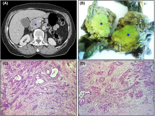
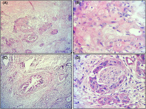
A 66-year-old previously healthy female patient presented with a one-month history of epigastric pain. Physical examination was remarkable for epigastric tenderness and jaundice. The serum tumor marker level for CA 19–9 was elevated to 975 U/ml (normal range = 38 U/ml). Computed tomography scan revealed an ill-defined and polylobed pancreatic head mass, with hypodense and heterogeneous enhancement (Figure 1A).1 The patient underwent pancreaticoduodenectomy. Macroscopically, we identified a poorly circumscribed greenish firm mass measuring 4.5 × 3.3 cm within the body of the pancreas (Figure 1B). Histologically, the tumor was composed of intimately admixed areas with squamous differentiation ≥30% and foci of ductal adenocarcinoma within a desmoplastic stroma (Figure 1C,D).2 The adenocarcinoma component was composed of infiltrating columnar cells showing glandular differentiation. The squamous component was composed of keratinizing neoplastic squamous cells arranged in nests or sheets of polygonal cells with distinct cell borders and eosinophilic cytoplasm and varying degrees of keratinization (Figure 2A,B). We identified low-grade pancreatic intraepithelial neoplasia (Figure 2C) and perineural invasion (Figure 2D). The tumor was confined within the pancreas, and 23 regional lymph nodes were not metastatic. The postoperative course was uneventful. Currently, the patient is systematically monitored on an outpatient basis with a follow-up period of 1 month.


AUTHOR CONTRIBUTIONS
Dr Faten LIMAIEM and Dr Mohamed HAJRI prepared, organized, wrote, and edited all aspects of the manuscript. Dr Faten LIMAIEM prepared all of the histology figures in the manuscript. Pr Leila BEN FARHAT and Dr Mohamed HAJRI participated in the conception and design of the study, the acquisition of data, analysis and interpretation of the data. All authors contributed equally to preparing the manuscript and participated in the final approval of the manuscript before its submission.
ACKNOWLEDGMENT
None.
FUNDING INFORMATION
The authors did not receive funding for this article.
CONFLICT OF INTEREST
None declared.
ETHICAL APPROVAL
All procedures performed were in accordance with the ethical standards. The examination was made in accordance with the approved principles.
CONSENT
Published with written consent of the patient.
Open Research
DATA AVAILABILITY STATEMENT
The data that support the findings of this study are available from the corresponding author upon reasonable request.


