Non-traumatic myositis ossificans circumscripta in the anterior abdominal wall of a seven-year-old Ugandan child: A case report
Abstract
Myositis ossificans circumscripta (MOC) is a benign and self-limiting heterotopic ossification in the subcutaneous fat, tendons, muscles, and nerves. It is commonly due to trauma and is frequently encountered in the arm, shoulder, thigh, and hand which are prone to trauma. Non-traumatic MOC arising from the abdominal muscles is extremely rare. We report a case of 7-year-old male child with a three-year history of progressive painless abdominal swelling in the left hypochondria region with no history of associated trauma. CT scan of the abdomen showed a well-defined hyperdense mass in the left external oblique muscle. Histological diagnosis confirmed myositis ossificans of the external oblique muscle. The mass was removed surgically with no immediate or late complications.
1 BACKGROUND
Myositis ossificans circumscripta (MOC) is a non-neoplastic, localized, and self-limiting soft-tissue tumor and is associated with prominent heterotrophic bone formation within the muscles, ligaments, and fascia.1 About 60 to 75% of all cases are due to trauma.2 MOC commonly affects young people but is rare in children and older patients.3 Men are more commonly affected than women during their second and third decades of life.3, 4
Though it can occur in any body part, MOC lesions are commonly found at sites most prone to injuries which are large muscles of the extremities.5 Myositis ossificans arising from the abdominal muscles is extremely rare, and if it appears, it tends to arise from abdominal operation scars.3, 6 MOC can be hereditary or non-hereditary.7, 8 The hereditary form (myositis ossificans progressiva) is due to an overexpression of bone morphogenetic protein 4(BMP4) mapped to chromosome 14.9, 10 The non-hereditary type is a commonly post-traumatic and well-defined lesion that usually complicates the hematoma of the affected muscles.2, 11 The signs and symptoms are specific and they can be confused with malignant lesions, such as osteosarcoma and soft-tissue sarcoma.12 Radiological imaging is crucial for excluding infections or malignancies and in the diagnosis of myositis ossificans.13 The diagnosis of myositis ossificans can be confirmed by histology.8, 14 Herein, we present histology confirmed non-traumatic MOC of the anterior abdominal wall in a seven-year-old Ugandan male.
2 CASE PRESENTATION
A 7-year-old male child presented to the Uganda cancer institute with a history of slowly progressive painless abdominal mass in the left hypochondria region. The mass had lasted for 3 years and was associated with a history of weight loss and drenching night sweats. There was no history of allergy or developmental abnormalities.
On general examination, he looked ill, mild pallor; however, not jaundiced, not febrile.
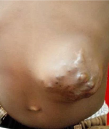
On physical examination, there was an irregular hard and non-tender mass measuring13x14cm. It was mobile and not attached to the underlying structures but attached to the underlying skin (Figure 1). There were no palpable regional lymph nodes or organomegaly.
Routine biochemical and hematological (Hb value 10 g/dL, WBC: 9000/mm3) investigations were carried out and were all within normal limits.
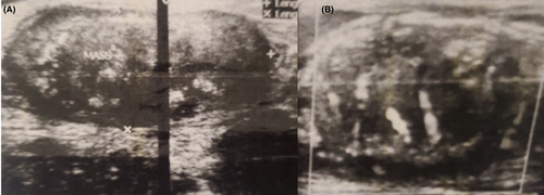
An abdominal ultrasound scan showed a subcutaneous solid mass in the left hypochondriac that was predominantly hypoechoic that measured 7 cm by 4 cm with acoustic shadowing in some areas. There was an increased flow on color Doppler. (Figure 2).
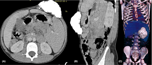
Computed tomography (CT) of the abdomen demonstrated a large micro-lobulated calcified hyperdense mass that measured 8 cm × 7cmx4cm in the left external oblique muscle abutting but not infiltrating the internal oblique muscle. The rest of the abdominal organs appear normal. There were no enlarged mesenteric lymph nodes (Figure 3).
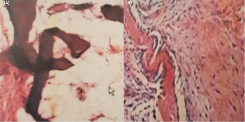
Under ultrasound guidance, trucut biopsies were taken and the sample was sent for histopathological evaluation. The hematoxylin and eosin-stained sections showed tissue composed of mature bone trabeculae, fibrous tissue, and muscle with foci of calcifications without malignant cells, which were features of MO (Figure 4). The mass was removed by surgical excision and no immediate or late complications were seen. There was no recurrence in a one-year follow-up.
3 DISCUSSION
MOC is a benign entity and yet it can mimic more aggressive pathology.15 It can be traumatic or non-traumatic.1 Some conditions share similar names or are related to MO—MO circumscripta, fibro-osseous pseudotumor of the digits, panniculitis ossificans, and MO progressiva (MOP).16 MOC occurs following traumas or surgical operations such as an abdominal scar, orthopedic operation, and dislocation.17 MOP mainly occurs in early life, progressively affects all skeletal muscles, and leads to death.18, 19 Non-traumatic MOC is the least common type and can easily be confused with malignant tumors, due to the lack of history of trauma.20 The presented case had no history of recorded or memorable trauma. The most common locations of MOC are the arm, shoulder, thigh, and hand,21 in our case, it arose from the left external oblique muscle, which is extremely rare.
MOC may occur at any age but rarely occurs in children and older patients.14, 22 The youngest documented case of MOC in the literature was a five-month-old girl22 and the oldest was an 81-year-old woman.14 Males are more commonly affected than females.4 The presented case was a 7-year-old male child.
Even though up to three-quarters of patients had a history of trauma, there are also non-traumatic MOC cases reported,23 which are similar to our case.
Radiological investigations are very important in the detection, diagnosis, and follow-up of MOC.18 Moreover, it is a useful tool for differentiating MOC from infections and malignancies.24 However, the radiological findings are typically vary depending on the lesion maturity.25, 26
MOC maturity has three phases which are active (early), subacute (intermediate), and chronic (mature).27 The presented case was in its mature or chronic phase during presentation.
Plain radiographs are mostly normal in the active phase and mineralization is seen in the chronic phase.13 Ultrasonography and magnetic resonance imaging (MRI) are important in the acute phase of symptomatology even before the appearance of calcifications.2, 13, 28 Ultrasound scan of the mass showed predominantly hypoechoic mass in the left hypochondriac with the increased flow on color Doppler.
The findings of myositis ossificans on MRI scans depend on the maturity phase.2, 24 MRI is not sensitive in soft-tissue calcification detection, as a result, it can be difficult to differentiate from adjacent muscles with marked surrounding edema in the early phases.12, 18 In the subacute phase, a hypointense border corresponding to peripheral calcification is observed, whereas the intensity decreases in the chronic phase.29
Computed tomography is the preferred imaging modality for detecting and diagnosing myositis ossificans since it is very good at showing the zonal patterns of the calcification that are expected at the different phases of MOC.20, 26 The differentials of MOC in the acute or subacute phases are muscle abscess, rhabdomyolysis, focal myositis, and soft-tissue sarcoma.12, 20 Bone scintigraphy is the very sensitive imaging modality in the early detection of myositis ossificans, shows increased uptake in damaged muscle.15 In the presented case, the lesion has lasted 3 years and the entire mass was made up of mature compact bone.
The CT scan revealed a large micro-lobulated calcified hyperdense mass in the left external oblique muscle of the abdomen.
Histopathology in the acute phase of some MOC cases could be misinterpreted for fibromatosis or sarcoma, but once it reaches the chronic phase, the diagnosis is clear.30 Our case was in the chronic phase, and histopathological examination showed tissue composed of mature bone trabeculae.
There are different management approaches reported in the literature, that includes immobilization, elevation, ice application, cold laser, acetic acid iontophoresis, extracorporeal shockwave therapy, and surgical excision.23, 31 Among the different management approaches, surgical excision is the most common treatment for non-traumatic myositis ossificans seen in an unusual site.32 The presented case had surgical excision of the mass.
4 CONCLUSION
Non-traumatic MOC of the abdominal muscles is extremely rare. Radiological investigations are very important in the diagnosis and necessary for differentiating a MO lesion from other malignant soft-tissue tumors.
AUTHOR CONTRIBUTIONS
HM, SG, VM, and ZM made substantial contributions to conception and design, acquisition of data, and interpretation of data; took part in drafting the article or revising it critically for important intellectual content. All agreed to submit to the current journal; gave final approval of the version to be published; and agree to be accountable for all aspects of the work.
ACKNOWLEDGMENTS
We would like to acknowledge the patient and patient's parent, the staff of the surgical oncology and radiology departments of Uganda cancer institute, for they actively supported the process of data collection and followed up updates of the patient.
CONFLICT OF INTEREST
The authors declare that they have no competing interests.
ETHICAL APPROVAL
No institutional approval was required to publish the case details. The parents of the child provided informed written consent to participate in the study of their child's condition.
CONSENT
The parents of the child provided informed written consent for this case to be published in a peer-reviewed journal.
Open Research
DATA AVAILABILITY STATEMENT
The data that support the findings of this study are openly available in [repository name e.g “figshare”] at https://doi.org/10.1002/ccr3.6145, reference number [reference number].


