Extra-gonadal mature teratoma: A case report of retroperitoneal location with a literature review
Abstract
Extra-gonadal mature teratoma is a benign tumor occurring rarely in adults. The retroperitoneal localization constitutes less than 4%. Treatment consists of surgical resection. Histological examination is essential for definitive diagnosis. We reported an unusual case of mature retroperitoneal teratoma discovered in a young man with abdominal pain.
1 INTRODUCTION
Teratomas are uncommon neoplasms that comprise a mixed dermal elements derived from the three germ cell layers.1, 2 They can be mature benign without malignant cells or immature with malignant cells.3 They commonly occur in the sacrococcygeal region in children and in gonads in adults especially in women. The retroperitoneal localization is not common. It constitutes less than 4% of all extragonadal teratomas.4 In addition, those benign tumors are most often asymptomatic. In most cases, they are identified incidentally.
In this paper, we report a case of mature extra-gonadal retroperitoneal teratoma occurring in a young adult man. We focus on clinical, radiological, biological, and histological presentations, and we realize a literature review of this rare location.
2 SEARCH STRATEGY
2.1 Publication search
We performed a literature search in PUBMED for English language sources using the following keywords chosen from the Medical Subject Headings (MeSH) of MEDLINE:(“teratoma”[Mesh]) AND (“retroperitoneal space”[Mesh]), (“dermoid cyst”[Mesh]) AND (“retroperitoneal space”[Mesh]). Only cases occurring in adults were included.
The search included studies published from the database inception to April 2022. A total number of 6 articles were included in this review article.
2.2 Collection data
The following data were collected from each case report: age, gender, symptoms, clinical examination, biologic findings, radiologic findings, intervention, histological findings, and outcomes.
The identified cases are shown in Table 1.3-8
| Reference | Age/sex (Year) | symptoms | Clinical findings | Laboratory findings | Imaging investigations | Surgery procedure | Histological findings | Outcomes |
|---|---|---|---|---|---|---|---|---|
| Shakir et al.3 | 31/female | Food indigestion, vomiting, abdominal pain | Palpable mass in left lumbar region | Normal |
Ultrasound: cystic mass in left hypochondrium CT scan: mass in left lumbar region anteromedial to left kidney |
Laparotomy with mass resection | Benign mature cystic teratoma | Simple |
| Lukanovic a et al.4 | 24/female | Pelvic pain and pressure | NS | NS | CT scan: mass in the left paravesical fossa altering the symmetry of the gluteal/perineal region | Laparotomy with mass resection | Benign mature cystic teratoma | Simple |
| Chen et al.5 | 50/female | Low back pain, night sweats | Normal | Normal |
CT scan: oval hypodense mass appearing arise from the left adrenal gland |
Laparoscopic exploration: resection of an independent mass |
Benign mature cystic teratoma |
Simple |
| Sherer et al.6 | 26/female | Pelvis pressure |
Mild left lower abdominal tenderness, deviated uterus with a posterolateral mass |
Normal |
Transvaginal ultrasound: encapsulated heterogeneous mass with debris CT scan: displaced vaginal vault and rectum by a large pelvic mass |
Laparotomy: the mass was opened, and the thick fluid contents of the cyst were removed by suction. |
Benign mature cystic teratoma | Simple |
| Talwar et al.7 |
23/ 32 weeks pregnant female |
Fever, upper abdominal pain | Tender mass was palpable in right upper abdomen | NS |
Ultrasound: hypoechoic mass suggestive of a large collection in subhepatic space CT scan: tumor with fluid, soft tissue, fat, and bone densities in the retroperitoneal space on right side |
Laparotomy: resection of a capsulated mass | Benign mature cystic teratoma with abscess |
Spontaneous labor simple |
| Kao et al.8 | 24/female | Low back pain with sciatica | Mild throbbing pain in the right flank | NS |
Lumbar spine radiograph: calcified tissues on the upper right quarter of the abdomen CT scan: delimited retroperitoneal mass, with extension into the intraspinal space at the L2-L4 level MRI: delineated intraspinal extradural cystic lesion |
Right subcostal incision: a mass compressing the kidney and ureter to the left with extension to L2–L3 neuroforamen into the posterior lateral epidural space hemilaminectomy with liberation and resection of the mass |
Benign mature cystic teratoma | Simple |
| Our case | 30/male | Left sided abdominal | Palpable oblong mass of 10 cm long axis in the left hypochondrium and the left flank | Normal | CT scan: heterogeneous retroperitoneal mass of the left flank exerting a mass effect on the left ureter, and responsible for a slight dilatation of excretory cavities | Midline laparotomy: resection of a left retroperitoneal mass blowing the left mesocolon | Benign mature cystic teratoma | Simple |
- Abbreviations: CT scan, computerized tomography scan; MRI, magnetic resonance imaging; NS, not specified.
3 CASE PRESENTATION
3.1 Case history and examination
Informed written consent was obtained from the patient before the publication of the case details and accompanying images.
A 30-years-old male patient, without previous comorbid, was referred to our surgery department for left sided abdominal pain evolving for 2 months. He had neither fever nor loss of weight and appetite. He had no digestive complaints or urinary symptoms.
The physical examination revealed an overweight, a normo-colored conjunctiva, a normal blood pressure, and a normal thermal profile. Cardiovascular, respiratory, and neurological examinations were normal. There was no testicular mass. On abdominal examination, there was a palpable oblong mass of 10 cm long axis in the left hypochondrium and in the left flank. It was deep, immobile on breathing, with firm consistency. Auscultation of bowel sounds was normal. Colonoscopy did not show any abnormality.
3.2 Differential diagnosis, investigations, and treatment
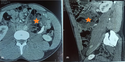
Abdominal computerizing tomography (CT) scan showed a heterogeneous retroperitoneal mass of the left flank, with mixed components of water, fat, and calcium (Figure 1). It measured 9 cm in long axis, exerting a mass effect on the left ureter, and responsible for a slight dilatation of excretory cavities. The chest X-ray was normal. There was no other suspected location.
The operability assessment was normal. World Health Organization performance status grade was 0. Hemoglobin level was 14.2 g/dl. Renal function was normal, and urines were sterile on cyto-bacterial examination.
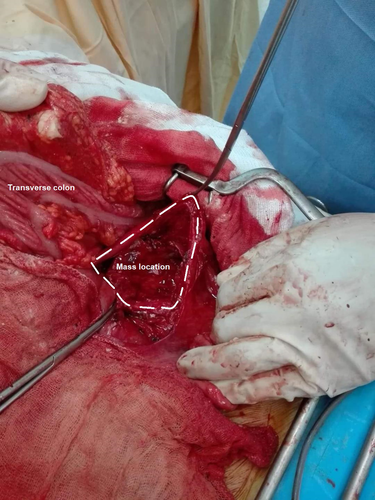
An exploratory midline laparotomy for mass resection was planified. Per-operative exploration showed a left retroperitoneal mass of 8 cm long axis, blowing the left mesocolon (Figure 2), and having a thick and inflammatory capsule. A careful left colic mobilization was performed after incision of the left mesocolon, in an avascular zone, in contact with the mass, from inside to outside. Gerota's fascia was intact. The mesocolic gap was sutured to avoid internal herniation later. The vascularity of the colon was not compromised. Enucleation of the mass was performed (Figure 3).
3.3 Outcome and follow-up
Post-operative course was simple. The patient was discharged home on the third postoperative day.
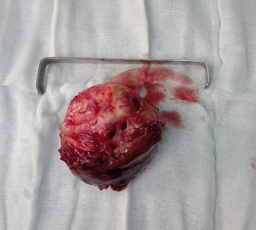
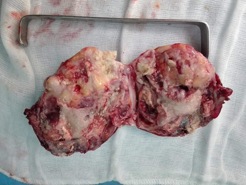
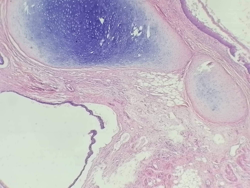
Pathological examination showed a heterogeneous mass on section, with adipose lobules, cartilaginous hair and mucus, and covered with false membranes (Figure 4). On microcopy, there were 3 types of epithelium; a keratinizing squamous epithelial, a respiratory type, and a secretory mucous gastric one with an extensive inflammatory remodeling (Figure 5). There was no immature component. The diagnosis of mature cystic teratoma was made. After 03 months of follow-up, the patient reported pain disappearance with a normal digestive transit.
4 DISCUSSION
Our case is of interest because it provides an overview of a rare extra-gonadal mature teratoma occurring in a young adult man in the retroperitoneal region.
Teratomas develop from the three embryonic germ layers.1 There are two variants: cystic teratomas which are composed of fully mature elements and are usually benign and solid teratomas which are more likely to be malignant.9, 10 Primary retroperitoneal teratomas constitute 1 to 11% of retroperitoneal neoplasms and are most commonly found in neonates and in young adults.2 They occur in female twice than male. Benign teratomas are usually asymptomatic.11 Symptoms are related to the increase in volume causing compression of surrounding structures .5, 12 As in our case, patient may present with back or abdominal pain, genito-urinary, or gastro-intestinal symptoms. Lower extremity or genital edema may be also noted.2 Additionally, some complications may happen, such as abscess or rupture causing peritonitis.7, 13
Physical examination may found a palpable abdominal mass, a tenderness, and distension.2, 14 This mass may be confused with other tumors such as renal cysts, adrenal tumors, gonadic tumors, fibromas, hemangiomas, enlarged lymph nodes, and sarcomas. Imaging investigations can help guide the diagnosis. Abdominal ultrasound can reveal tumors with cystic and solid components and echogenic spots with acoustic shadows with some limitations in detecting fat and calcifications.14 Hence, CT scan offers a better identification of fat, sebum, and adipose tissue. Therefore, identification of sebum on CT is very suggestive for teratoma.2 However, the diagnostic accuracy remains histological.8, 15 Surgical resection is of greatest importance for both diagnosis and treatment. Midline or transverse incision is used for most patients. When possible, laparoscopic methods may be an excellent alternative but requires an adequate laparoscopic skills.16 Usually, primary retroperitoneal teratoma does not infiltrate adjacent structures and can be fully resected without significant complications. Prognosis is excellent in patients with complete resection of benign teratomas. Malignant transformation of mature teratoma is uncommon. Also, malignant teratomas with germ cell elements may have a grim prognosis, hence, the interest of a complete surgical resection with a histological confirmation.17
Through our literature review, we came across 6 cases of primary mature extra-gonadal retroperitoneal teratomra occurring in adult patients. They are presented in Table 1.3-8 The age of patients ranged from 23 to 50 years with an average of 29 years. They were 6 females. Abdominal pain was the most common first clinical presentation. Abdominal scan was performed for all patients. The mass was localized in the left retroperitoneal region for most of them. Complications were noted in two cases, one with abscess, and one with intra spinal extension.
Regarding therapy, laparotomy was performed in 5 cases and laparoscopy in one case. Simple outcome was noted in all cases.
5 CONCLUSION
Primary retroperitoneal mature teratomas with extra-gonadal location are rare especially in young adult man. They are usually identified when they exert a mass effect on adjacent structures. Surgical resection remains the mainstay of therapy, and histological examination is essential for definitive diagnosis. Patients with complete resection of a benign teratoma have an excellent prognosis.
Additionally, we have highlighted teratoma as a differential diagnosis of retroperitoneal mass, and we emphasized the importance of complete resection to confirm the diagnosis and to avoid complications and malignancy transformation.
AUTHOR CONTRIBUTIONS
All the authors participated in the design of the study and wrote the manuscript. All authors read and approved the final manuscript. Mohamed Farès Mahjoubi has drafted the work. Ghassen Hamdi Kbir has substantively revised the work. Mohamed Maatouk, Sohaib Messaoudi, and Bochra Rezgui have made substantial contributions to the literature research. Mounir Ben Moussa has made substantial contributions to the conception of the work.
ACKNOWLEDGMENTS
None.
CONFLICT OF INTEREST
The authors have no conflict of interests.
ETHICAL APPROVAL
Our locally appointed ethics committee “Charles Nicolle Hospital local committee” has approved the research protocol and informed consent has been obtained from the subject.
HUMAN AND ANIMAL RIGHTS
Our study complies with the Declaration of Helsinki.
CONSENT
Written informed consent was obtained from the patient.
Open Research
DATA AVAILABILITY STATEMENT
The data and supportive information are available within the article.


